
Figures & data
Figure 1. Omentin protected PC12 cell death against H2O2-induced apoptosis. (A,B) The effect of omentin on the viability of PC12 cells. The cells were incubated with different concentrations of omentin (100, 200, 500, and 1000 ng/mL) for 24 h, 1000 ng/mL omentin for different time (1, 2, 5, 12, and 24 h), respectively. (C–F) The protective effect of omentin on H2O2-induced injury. After preincubation with different dose of omentin for 24 h, PC12 cells were treated with 100 μM H2O2 for 4 h. Cell viability was assessed by CCK8 (C), and cell apoptosis rates were assessed by TUNEL assays (D–E) and flow cytometry (F). Data are expressed as mean ± SD (n = 3) (*p<.05; **p<.01).
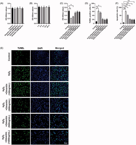
Figure 2. Omentin inhibited the apoptosis of PC12 cells induced by H2O2. (A) PC12 cells were pre-incubated with 500 ng/mL omentin for 24 h before exposure to H2O2 for 4 h. The antioxidant enzymes of superoxide dismutase (SOD), catalase (CAT), glutathione peroxidase (GSH-Px), and malondialdehyde (MDA) were analyzed. (B) The western blotting assay was used to detect the expression levels of proteins related to apoptosis, including Bcl-2, Bax, pro-Caspase3, and cleaved-Caspase3, while β-actin was utilized as an internal control. (C) PC12 cells were cultured with different doses of omentin for 24 h, and the western blotting assay was used to detect the expression levels of proteins related to apoptosis. Results are presented as mean ± SD (n = 3) (*p<.05; **p<.01).
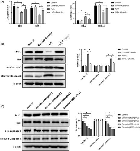
Figure 3. MiR-128-3p suppresses Bax expression. (A) Bioinformatics analysis of microRNA upstream of Bax. (B) The expression levels of different miRNAs in PC12 cells co-cultured with 500 ng/mL omentin. (C) The expression levels of different miRNAs in PC12 cells pre-incubated with 500 ng/mL omentin before exposed to H2O2. (D–G) The effect of miR-128-3p mimics (D & F) and inhibitor (E & G) on the Bax expression was detected by RT-qPCR (D & E) and Western blotting assay (F & G), respectively. (H) Luciferase assay of HEK 293 T co-transfected with wild-type (WT) or mutated (MUT) Bax 3′-UTR and miR-128-3p mimics. Results are presented as mean ± SD (n = 3) (*p<.05; **p<.01).
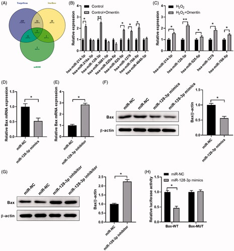
Figure 4. MiR-128-3p mimics possess the potential effect against H2O2-induced apoptosis. (A) The changes in miR-128-3p expression levels transfected with miR-128-3p mimics followed by incubating with H2O2. (B–D) The cell viability of PC12 cells treated with H2O2 and miR-128-3p mimics was analyzed by CCK8 (B), and the cell apoptosis rates were detected by TUNEL assay (C,D). (E) The cell apoptosis rates were detected by flow cytometry. (F) The effect of H2O2 and miR-128-3p mimics on apoptosis-related proteins expression were detected by western blotting assay. Results were presented as mean ± SD (n = 3) (*p<.05, **p<.01).
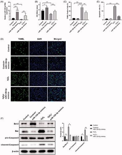
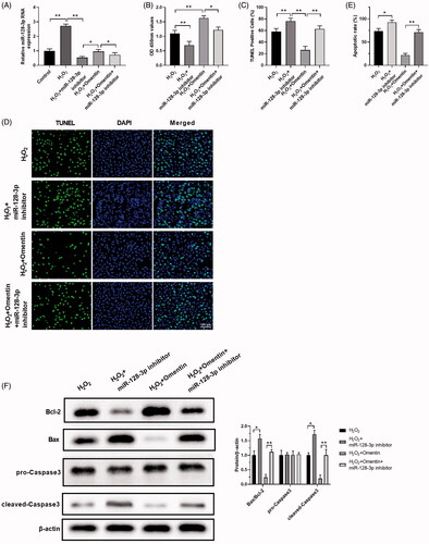
Figure 5. MiR-128-3p inhibitors alleviate the protective effect of omentin against H2O2-induced apoptosis. (A) The changes of miR-128-3p expression level after treated with H2O2, H2O2 + miR-128-3p inhibitor, H2O2 + omentin, H2O2 + omentin + miR-128-3p inhibitor. (B–D) The cell viability of PC12 cells in different groups were detected by CCK8 assay (B), and the cell apoptosis rates were detected by TUNEL assay (C,D). (E) The cell apoptosis rates were detected by flow cytometry. (F) The effect of H2O2, miR-128-3p inhibitor, and omentin on the expression of proteins related to apoptosis were detected by Western blotting assay. Results were presented as mean ± SD (n = 3) (*p<.05; **p<.01).
Supplemental Material
Download TIFF Image (307.1 KB)Data availability statement
The datasets generated and/or analyzed during the present study are available from the corresponding author upon reasonable request.