
Figures & data
Figure 1. Effects of LIPUS on the body weight, grip strength, and muscle weight in CKD mouse models. Two mouse models of unilateral IRI with contralateral nephrectomy (1) and adenine administration (2) were used. Body weight (A), grip strength for forelimb and hindlimb (B), and muscle weights for gastrocnemius, tibialis anterior, and soleus muscles (C) were shown. Data are presented as mean ± SEM (n = 8 for IRI model; n = 9 for adenine model). *p < 0.05, versus sham/control group; #p < 0.05, versus CKD alone group.
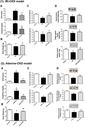
Figure 2. Effects of LIPUS on the cross-section areas of myofibers and fibrosis in soleus muscles of CKD mouse models. Two mouse models of unilateral IRI with contralateral nephrectomy (1) and adenine administration (2) were used. Cross-section areas (CSA) of myofibers (A-a), quantification of CSA (A-b), and distribution of myofibers (A-c) in soleus muscles were shown. Moreover, the collagen depositions in soleus muscles of IRI-CKD mice were stained by Masson’s Trichrome (B). The quantification was shown. Data are presented as mean ± SEM (n = 8 for IRI model; n = 9 for adenine model). *p < 0.05, versus sham group; #p < 0.05, versus CKD alone group.
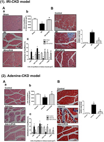
Figure 3. Effects of LIPUS on the immunointensity for phosphorylated Akt, Atrogin-1, and MuRF1 in soleus muscles of CKD mouse models. Two mouse models of unilateral IRI with contralateral nephrectomy (1) and adenine administration (2) were used. Immunohistochemistry for the protein expression of phosphorylated Akt, Atrogin-1, and MuRF1 was shown. The relative optical density of immunohistochemical images in five random visual fields of each section was determined by the image J 1.48 software. Data are presented as mean ± SEM (n = 8 for IRI model; n = 9 for adenine model). *p < 0.05 versus sham group; #p < 0.05 versus CKD alone group.
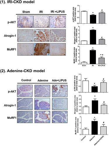
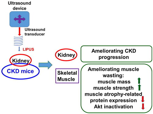
Figure 4. A schematic summary of our main findings for the effects of LIPUS on CKD-associated muscle wasting.
Supplemental Material
Download TIFF Image (7.2 MB)Supplemental Material
Download TIFF Image (7.3 MB)Supplemental Material
Download TIFF Image (6.9 MB)Supplemental Material
Download TIFF Image (3.1 MB)Supplemental Material
Download PDF (281.3 KB)Supplemental Material
Download TIFF Image (7.5 MB)Supplemental Material
Download TIFF Image (6.2 MB)Supplemental Material
Download TIFF Image (3.3 MB)Data availability statement
All data supporting the findings of this study are included in the article (and its Supplementary Information files).