
Figures & data
Figure 1. V/sAS/Ag and V/pAS/Ag Fabrication processes and surface micromorphology of each material. A: Schematic of the construction of a silver HTZ sample downloaded under vacuum and non-vacuum conditions, with the base ceramic sheet in grey, the aluminosilicate coating in red, and the silver particles in particles. B: Representative images of sintered materials. C: Representative images of observation of the surface morphology of different materials.
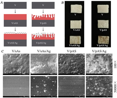
Figure 2. EDS elemental analysis of Material surfaces. A: Representative images of V/sAS/Ag element proportion and spectrum; B: Representative images of V/pAS/Ag element proportion and spectrum.
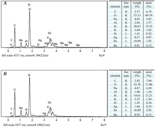
Figure 3. XPS detection of Material surface elements. A: Representative images of full spectrum of vacuum sintered unloaded silver sample V; B: Representative images of full spectrum of vacuum sintered coated V/sAS/Ag sample. C: Representative images of full spectrum of non-vacuum sintered coated V/pAS/Ag sample.
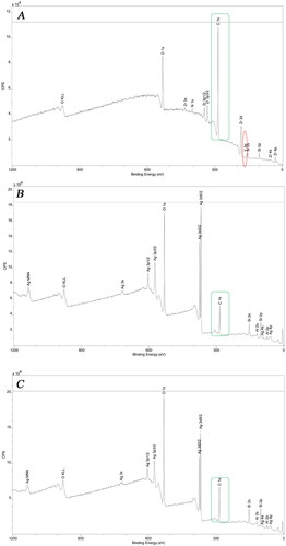
Figure 4. Silver element detection. A: Representative images of transmittance of modified V/sAS/Ag and V/pAS/Ag; B: Representative images of V/sAS/Ag and V/pAS/Ag silver particle distribution; C: Silver ion release curves in V/sAS/Ag and V/pAS/Ag. T-test was used for expression differences between the two groups, * p < 0.05.
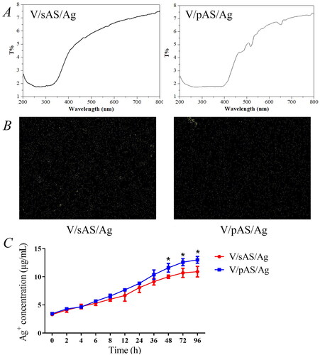
Figure 5. Size of inhibition zone around V/sAS/Ag and V/pAS/Ag.
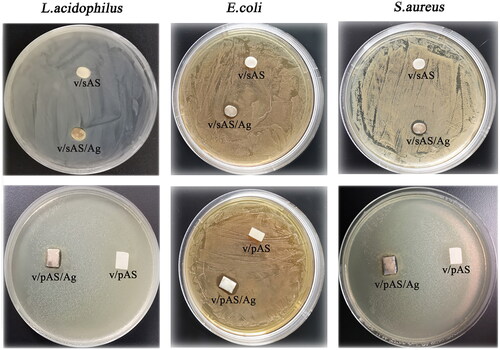
Figure 6. The sterilisation effect of silver-loaded materials. A: Bactericidal effect of V/sAS/Ag and V/pAS/Ag treatment on S.aureus, L.acidophilus, and E.coli. Statistical plots of the bactericidal effects of V/sAS/Ag and V/pAS/Ag on S.aureus (B), L. acidophilus (C), and E.coli (D). compared with the control group, **p < 0.01; compared with the V/sAS/Ag group, #p < 0.05.
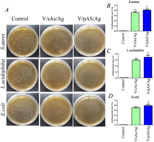
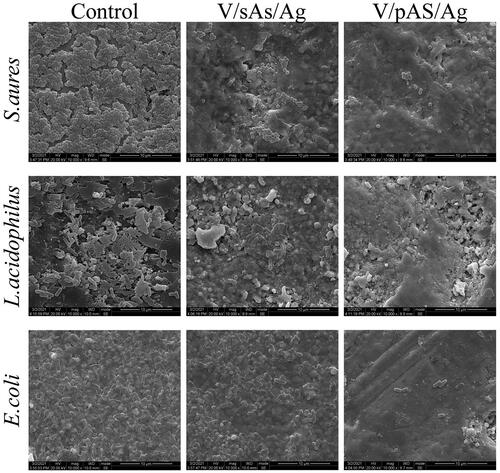
Figure 7. Representative SEM images of S.aureus, L.acidophilus, and E.coli after the action of V/sAS/Ag and V/pAS/Ag.
Data availability statement
The datasets used or analyzed during the current study are available from the corresponding author on reasonable request.