2,347
Views
24
CrossRef citations to date
0
Altmetric
Full Length Articles

Porous structure engineering of bioceramic hydroxyapatite-based scaffolds using PVA, PVP, and PEO as polymeric porogens

ORCID Icon &
Pages 161-169 | Received 13 Oct 2018, Accepted 09 Feb 2019, Published online: 30 Mar 2019

Figures & data

Figure 1. FTIR spectra of: (a) raw materials of golden apple snail (Pomacea canaliculata) and (b) decomposed materials after calcination at high temperatures.
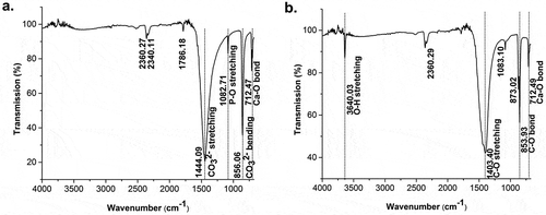
Figure 1. FTIR spectra of: (a) raw materials of golden apple snail (Pomacea canaliculata) and (b) decomposed materials after calcination at high temperatures.

Figure 2. Morphological changes of the precursors to form HAp: (a) raw golden apple snail (Pomacea canaliculata) shell; (b) CaO decomposed from raw materials; (c) HAp after sintering at 1000°C for 6 h; and (d) HAp powder.
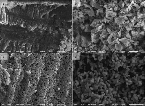
Figure 2. Morphological changes of the precursors to form HAp: (a) raw golden apple snail (Pomacea canaliculata) shell; (b) CaO decomposed from raw materials; (c) HAp after sintering at 1000°C for 6 h; and (d) HAp powder.

Figure 3. FTIR spectrum of synthesized HAp powder.

Figure 3. FTIR spectrum of synthesized HAp powder.

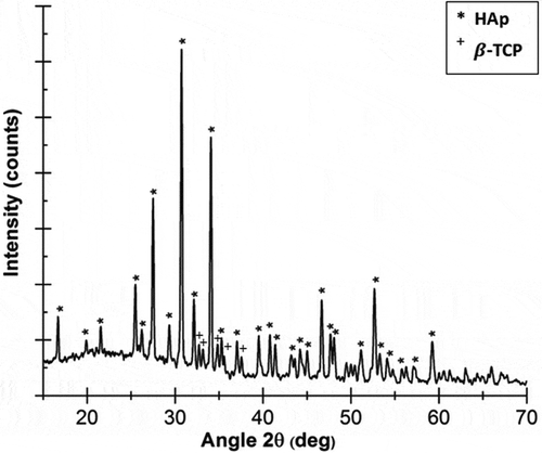
Figure 4. X-ray pattern diffraction of synthesized HAp.

Figure 4. X-ray pattern diffraction of synthesized HAp.

Figure 5. Increases in the pore size of HAp scaffolds as the porogen (PVA, PVP, or PEO) concentration is increased.
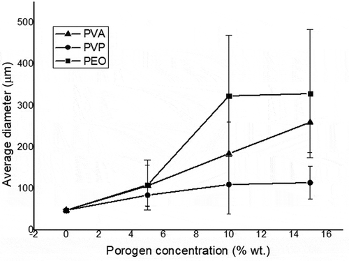
Figure 5. Increases in the pore size of HAp scaffolds as the porogen (PVA, PVP, or PEO) concentration is increased.

Figure 6. Macropore morphologies of hydroxyapatite (HAp) scaffolds fabricated using polyvinyl alcohol (PVA) as a porogen in the following concentrations: (a) 0 wt %; (b) 5 wt %; (c) 10 wt %; and (d) 15 wt % (Insets: HAp-based scaffolds magnified 3000 times).
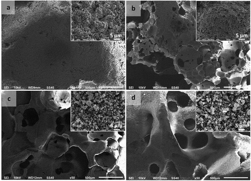
Figure 6. Macropore morphologies of hydroxyapatite (HAp) scaffolds fabricated using polyvinyl alcohol (PVA) as a porogen in the following concentrations: (a) 0 wt %; (b) 5 wt %; (c) 10 wt %; and (d) 15 wt % (Insets: HAp-based scaffolds magnified 3000 times).
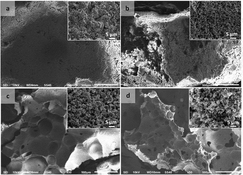
Figure 7. Macropore morphologies of hydroxyapatite (HAp) scaffolds fabricated using polyvinylpyrrolidone (PVP) as a porogen in the following concentrations: (a) 0 wt %; (b) 5 wt %; (c) 10 wt %; and (d) 15 wt % (Insets: HAp-based scaffolds magnified 3000 times).
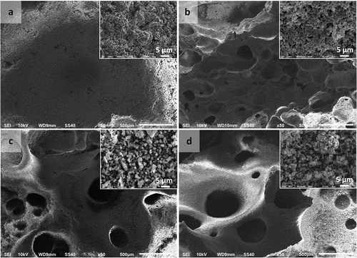
Figure 7. Macropore morphologies of hydroxyapatite (HAp) scaffolds fabricated using polyvinylpyrrolidone (PVP) as a porogen in the following concentrations: (a) 0 wt %; (b) 5 wt %; (c) 10 wt %; and (d) 15 wt % (Insets: HAp-based scaffolds magnified 3000 times).

Figure 8. Macropore morphologies of hydroxyapatite (HAp) scaffolds fabricated using polyethylene oxide (PEO) as a porogen in the following concentrations: (a) 0 wt %; (b) 5 wt %; (c) 10 wt %; and (d) 15 wt % (Insets: HAp-based scaffolds magnified 3000 times).

Figure 8. Macropore morphologies of hydroxyapatite (HAp) scaffolds fabricated using polyethylene oxide (PEO) as a porogen in the following concentrations: (a) 0 wt %; (b) 5 wt %; (c) 10 wt %; and (d) 15 wt % (Insets: HAp-based scaffolds magnified 3000 times).

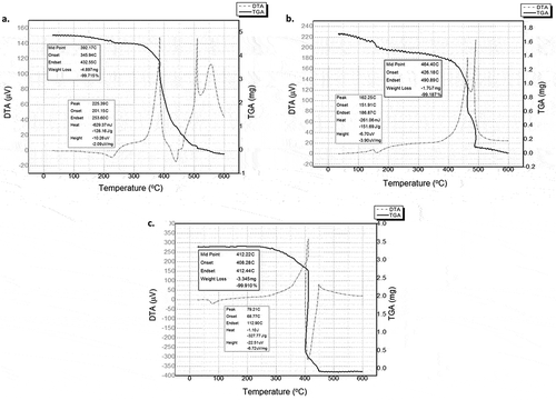
Figure 9. Thermal analysis of the porogens: (a) PVA; (b) PVP; and (c) PEO.

Figure 9. Thermal analysis of the porogens: (a) PVA; (b) PVP; and (c) PEO.

Figure 10. FTIR spectra of HAp-based scaffolds fabricated using porogens: (a) PVA; (b) PVP; and (c) PEO.
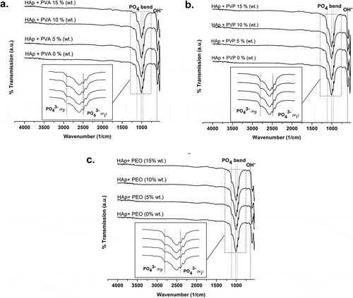
Figure 10. FTIR spectra of HAp-based scaffolds fabricated using porogens: (a) PVA; (b) PVP; and (c) PEO.
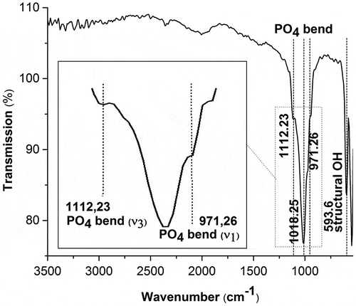
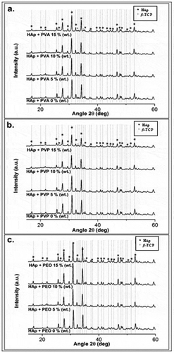
Figure 11. X-ray diffraction patterns of HAp-based scaffolds fabricated using (a) PVA; (b) PVP; and (c) PEO.

Figure 11. X-ray diffraction patterns of HAp-based scaffolds fabricated using (a) PVA; (b) PVP; and (c) PEO.

Figure 12. Effects of porogen concentration on HAp scaffold microhardness: (a) PVA; (b) PVP; and (c) PEO.
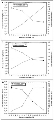
Figure 12. Effects of porogen concentration on HAp scaffold microhardness: (a) PVA; (b) PVP; and (c) PEO.