Figures & data
Figure 1. Geographical distribution of Bactrian camels. Geographical distribution of dromedary camels, Bactrian camels and wild Bactrian camels, including the area in which dromedary camels and Bactrian camels are co-localized.
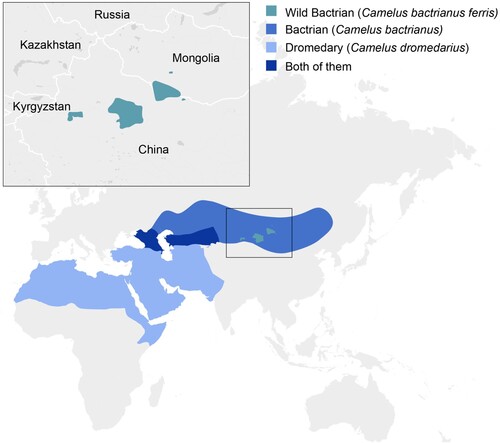
Figure 2. Bactrian camel dipeptidyl peptidase 4 (DPP4) analyses. (A) DPP4 amino acid sequence identity matrix. White indicates no similarity, dark red indicates 100% similarity. (B) DPP4 species amino acid variation within the 14 described contact points with MERS-CoV spike. (C) Co-structure of MERS-CoV spike interaction with human DPP4 (PDB: 4L72). Inset shows a top-down view of the 14 spike-contact points on DPP4 (coloured in red). (D) Co-structure of MERS-CoV spike interaction with the predicted structure of Bactrian camel DPP4 (Swissmodel). Inset shows a top-down view of the 14 spike-contact points on DPP4 (coloured in red).
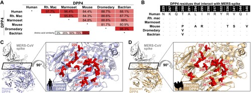
Figure 3. Clinical signs in Bactrian camels inoculated with Middle East respiratory syndrome coronavirus (MERS-CoV). (A) Nasal discharge observed in Bactrian camel 2; both Bactrian camels displayed nasal discharge during the experiment (B) Rectal temperature during the experiment. Rectal temperatures are indicated for each camel by lines with geometric shapes.
Figure 4. MERS-CoV shedding and tissue distribution in Bactrian camels. (A) Virus shedding from the upper respiratory tract in Bactrian camels inoculated with MERS-CoV determined by plaque assay. (B,C) Replication of MERS-CoV in the upper respiratory tract of Bactrian camels determined by qRT-PCR and plaque assay. The dotted line indicates the detection limits of the assays.
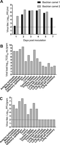
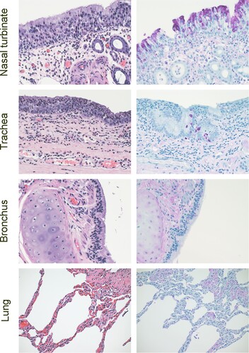
Figure 5. Histopathology in Bactrian camels infected with MERS-CoV. Histopathologic changes at 5 days post inoculation in camel 2 inoculated with MERS-CoV. Tissues were collected and stained with hematoxylin and eosin (left panel). AntiMERS-CoV immunohistochemical results (right panel) are visible as a dark-purple stain. Viral antigen was detected within the epithelial cells of the nasal turbinates (primarily neuroeptithelium) and trachea (columnar epithelium). Original magnification ×400.