Figures & data
Figure 1. 9F4 displays broad binding affinity and neutralizing activity against H5Nx viruses. (A) Alignment of residues 50–80 in the HA protein of H5Nx viruses with the corresponding domain in one clade 1 (VN04) and three other clade 2.3.4.4 viruses (Iowa15, GD14 and Korea14). For comparison, H3 numbering is followed. The conservation of R62, W69 and F79, which are important for the interaction with 9F4, are boxed. (B) ELISA was performed to determine the ability of 9F4 to bind to H5Nx-HA. Wells were coated with HA proteins of H5Nx (H5N1 (A/Vietnam/1194/2004); H5N2 (A/chicken/Iowa/04-20/2015); H5N6 (A/duck/Guangdong/GD01/2014); H5N8 (A/broiler duck/Korea/Buan2/2014)) and incubated with serially diluted 9F4. 1A4 was also used at the highest concentration (50 ng/ml) as an isotype control antibody. (C) Pseudotyped lentiviral particles harbouring the HA proteins of H5Nx IAV were incubated with 10-fold serially diluted 9F4-WT for 1 h at RT before inoculation onto MDCK cells. Luciferase activity in the cell lysates was determined at 48 hpi. Viral entry, as indicated by the luciferase activity, was expressed as a percentage of the reading obtained in the absence of antibody (No Ab), which was set at 100%. 1A4 was also used at the highest concentration (10 μg/ml) as an isotype control antibody. Representative data from three independent experiments are shown and error bars represent standard error of the mean (SEM) of the experiment carried out in triplicates.
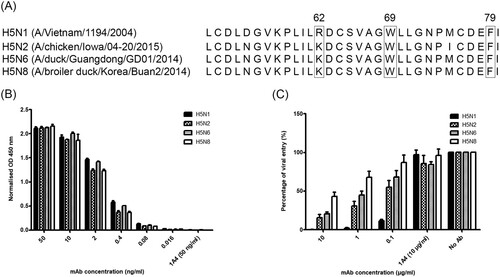
Figure 2. 9F4-WT and its Fc mutants display similar binding affinities to H5N6-HA protein and neutralize against H5N6 pseudotyped particles. ELISA was performed to compare the binding affinity of 9F4-WT with (A) 9F4-LALA or (B) 9F4-K322A to H5N6-HA. Wells were coated with H5N6-HA protein and incubated with serially diluted mAbs as indicated. 1A4 was also used at the highest concentration (50 ng/ml) as an isotype control antibody. Pseudotyped lentiviral particles harbouring the HA proteins of H5N6 IAV were incubated with 10-fold serially diluted 9F4-WT, (C) 9F4-LALA or (D) 9F4-K322A for 1 h at RT before inoculation onto MDCK cells. Luciferase activity was determined as above. Representative data from three independent experiments are shown and error bars represent SEM of the experiment carried out in triplicates.
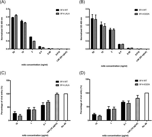
Figure 3. mFcγRIV activation is induced by 9F4-WT and 9F4-K322A but not 9F4-LALA. Mouse FcγRIV-based ADCC reporter assay was performed for 9F4-WT, (A) 9F4-LALA and (B) 9F4-K322A as described in materials and methods. 1A4 was used as an isotype control. Representative data of three independent experiments carried out in triplicates are shown and error bars represent SEM between replicates. Symbols represent statistically significant differences at p < 0.05 when comparing 9F4-WT with its Fc mutants (red asterisk) or 1A4 (blue hash), or Fc mutants with 1A4 (green carat). (C) Binding of human C1q to 9F4-K322A is abolished. C1q binding ELISA was performed to compare the binding affinity of human recombinant C1q to 9F4-WT and its Fc mutants. Wells were coated with H5N6-HA protein and incubated with serially diluted mAbs, followed by the addition of c1q protein. 1A4 was also used at the highest concentration (1 μg/ml) as an isotype control antibody. Representative data from three independent experiments are shown and error bars represent SEM of the experiment carried out in triplicates. Symbols represent statistically significant difference at p < 0.05 when compared to 9F4-WT (red asterisk) or 9F4-LALA (blue hash).
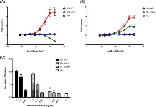
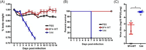
Figure 4. ADCC and/or ADCP contributes to the in vivo protective function of 9F4. Balb/c mice were infected with 100 PFU of rgPR8 H5N6 and received a single therapeutic dose of 9F4-WT, 9F4-LALA or the isotype control 1A4 at 10 mg/kg via intraperitoneal injections 24 hpi. (A) Body weight profiles and (B) animal survival are shown. (C) Lung virus titres were determined at 4 dpi by plaque assays with MDCK cells. n = 8 per treatment group, error bars represent SEM, asterisks indicate p < 0.05. (D) Representative images of H&E stained formalin fixed paraffin embedded lung sections along with their (E) corresponding histopathological grades of individual mice and averages of treatment groups at 4 or 14 dpi are shown.
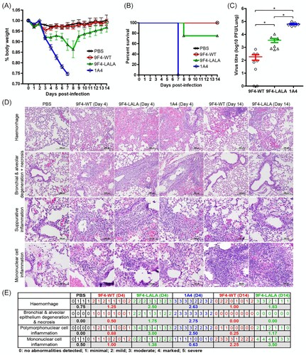
Figure 5. CDC is dispensable for the in vivo protective function of 9F4. Balb/c mice were infected with 100 PFU of rgPR8 H5N6 and received a single therapeutic dose of 9F4-WT, 9F4-K322A or the isotype control 1A4 at 10 mg/kg via intraperitoneal injections 24 hpi. (A) Body weight profiles and (B) animal survival are shown. n = 4. (C) Lung virus titres were determined at 4 dpi by plaque assays with MDCK cells. n = 8 per treatment group. Error bars represent SEM, asterisks indicate p < 0.05. (D) Representative images of H&E stained formalin fixed paraffin embedded lung sections along with their (E) corresponding histopathological grades of individual mice and averages of treatment groups at 4 dpi. n = 4 per treatment group.
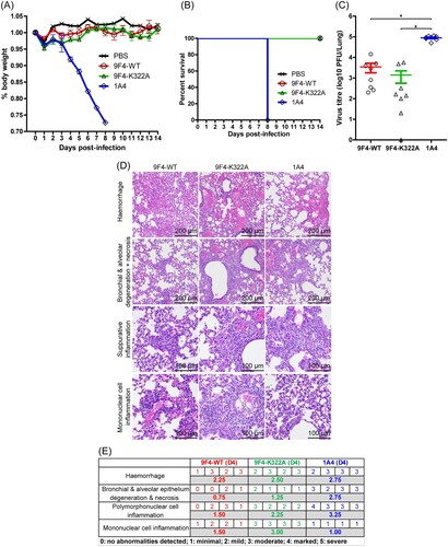
Figure 6. Alveolar macrophages are crucial for in vivo protection against H5N6 IAV infection by 9F4-WT. Balb/c mice were intranasally given 20 µl of clodronate encapsulated liposomes (CL) or control liposomes (PBS) 2 days prior and simultaneously during infection with 100 PFU of rgPR8 H5N6. Infected mice received a single therapeutic dose of 9F4-WT or the isotype control 1A4 at 10 mg/kg whereas mock-infected mice received an equivalent volume of PBS via intraperitoneal injections 24 hpi. (A) Body weight profiles and (B) animal survival are shown. (C) Lung virus titres were determined at 4 dpi by plaque assays with MDCK cells. (D) Representative images of H&E stained formalin fixed paraffin embedded lung sections along with their (E) corresponding histopathological grades of individual mice and averages of treatment groups at 4 dpi. n = 4 per treatment group. Error bars represent SEM, asterisks indicate p < 0.05.
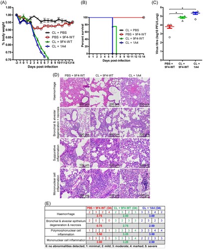
Figure 7. Intranasal administration of 9F4-WT is able to protect against H5N6 challenge in vivo. Balb/c mice were infected with 100 PFU of rgPR8 H5N6 and received a single therapeutic dose of 9F4-WT, 9F4-LALA or the isotype control 1A4 at 2 mg/kg via intranasal instillations 24 hpi. (A) Weight loss profiles and (B) animal survival are shown. (C) Lung virus titres were determined 4 dpi by plaque assays with MDCK cells. n = 4 per treatment group, error bars represent SEM, asterisks indicate p < 0.05.