Figures & data
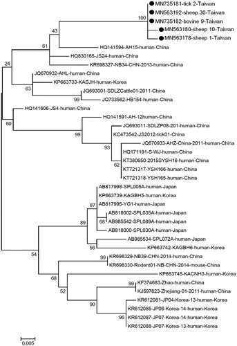
Figure 1. Phylogenetic analysis of the partial S segment of severe fever with thrombocytopenia syndrome virus identified in Taiwan. In total, of those 21 positive samples, 5 distinct sequences of partial S gene were identified from each species of animals in this study. The Nucleotide sequences of the local isolate indicated as a black circle. Other representative viral strains were presented with their accession numbers and also the host and country of isolation. The evolutionary history was inferred using the maximum-likelihood method, based on the Kimura 2-parameter model (1000 bootstrap replicates). The percentage of trees in which associated taxa clustered is shown next to the branches. Scale bar indicates nucleotide substitutions per position.