Figures & data
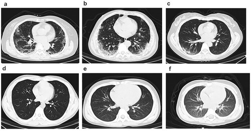
Figure 1. Chest CT images. (a) Transverse chest CT images from Case 1 showing bilateral ground-glass opacity, subsegmental areas of consolidation and subpleural line. (b) Transverse chest CT images from Case 2 showing peripheral pulmonary parenchymal ground-glass and consolidative pulmonary opacities. (c) Transverse chest CT images from Case 3 showing subsegmental areas of ground-glass opacity and consolidation. Transverse chest CT images from Case 4 (d), Case 5 (e) and Case 6 (f) were normal.