23,750
Views
318
CrossRef citations to date
0
Altmetric
Review

Colistin and its role in the Era of antibiotic resistance: an extended review (2000–2019)

ORCID Icon, , ORCID Icon, , &
Pages 868-885 | Received 10 Jul 2019, Accepted 04 Apr 2020, Published online: 06 May 2020

Figures & data

Figure 1. (a) Structures of colistin A and B; (b) structures of sodium colistin A and B methanesulphonate. Fatty acid: 6-methyl-octanoic acid for colistin A and 6-methyl-heptanoic acid for colistin B; Thr: threonine; Leu: leucine; Dab: α, γ-diaminobutyric acid. α and γ indicate the respective amino groups involved in the peptide linkage. Adapted from Li et al. [Citation16].

Figure 1. (a) Structures of colistin A and B; (b) structures of sodium colistin A and B methanesulphonate. Fatty acid: 6-methyl-octanoic acid for colistin A and 6-methyl-heptanoic acid for colistin B; Thr: threonine; Leu: leucine; Dab: α, γ-diaminobutyric acid. α and γ indicate the respective amino groups involved in the peptide linkage. Adapted from Li et al. [Citation16].

Figure 2. PRISMA-modified flow diagram of included and excluded studies. Adapted from the PRISMA website (http://www.prisma-statement.org/PRISMAStatement/FlowDiagram) and Liberati et al. [Citation11].

Figure 2. PRISMA-modified flow diagram of included and excluded studies. Adapted from the PRISMA website (http://www.prisma-statement.org/PRISMAStatement/FlowDiagram) and Liberati et al. [Citation11].

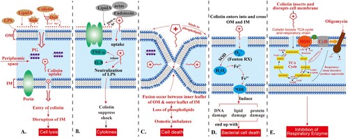
Figure 3. Action of colistin on the Gram-negative bacterial membrane. The cationic cyclic decapeptide structure of colistin binds with the anionic LPS molecules by displacing Mg2+ and Ca2+ from the outer cell membrane of Gram-negative bacteria, leading to permeability changes in the cell envelope and leakage of cell contents. LPS: lipopolysaccharides; PG: peptidoglycan; Dab: diaminobutyric acid (Dab); OM: outer membrane; IM: inner membrane. The scheme shows the five different mechanisms of antibacterial activity of colistin, namely; (A) Direct antibacterial colistin activity: the initial fusion of colistin with the bacterial membrane occurs via electrostatic interactions between the cationic diaminobutyric acid (Dab) residues of colistin and anionic phosphate groups on the lipid A moiety of LPS in the outer membrane, thus disrupting the bacterial outer and inner membranes and leads to cell lysis; (B) Anti-endotoxin colistin activity: The lipid A portion of LPS represents an endotoxin in Gram-negative bacteria. Thus, colistin inhibits the endotoxin activity of lipid A by binding to and neutralizing the LPS molecules, thus suppress the induction of shock through the release of cytokines such as tumour necrosis factor-alpha (TNF-α) and Interleukin 8 (IL-8); (C) Vesicle-Vesicle contact pathway: colistin bind to anionic phospholipid vesicles after transiting the OM leads to the fusion of the inner leaflet of the outer membrane with the outer leaflet of the cytoplasmic membrane, leading to loss of phospholipids and cell death; (D) Hydroxyl radical death pathway: Colistin acts via the production of the reactive oxygen species (ROS) this is known as, Fenton reaction, causing damage of DNA, lipid, and protein, and end up with cell death; and (E) Inhibition of respiratory enzymes: the antibacterial colistin activity is via the inhibition of the vital respiratory enzymes. Figure created using Adobe Illustrator version CC 2019 (23.1.0).

Figure 3. Action of colistin on the Gram-negative bacterial membrane. The cationic cyclic decapeptide structure of colistin binds with the anionic LPS molecules by displacing Mg2+ and Ca2+ from the outer cell membrane of Gram-negative bacteria, leading to permeability changes in the cell envelope and leakage of cell contents. LPS: lipopolysaccharides; PG: peptidoglycan; Dab: diaminobutyric acid (Dab); OM: outer membrane; IM: inner membrane. The scheme shows the five different mechanisms of antibacterial activity of colistin, namely; (A) Direct antibacterial colistin activity: the initial fusion of colistin with the bacterial membrane occurs via electrostatic interactions between the cationic diaminobutyric acid (Dab) residues of colistin and anionic phosphate groups on the lipid A moiety of LPS in the outer membrane, thus disrupting the bacterial outer and inner membranes and leads to cell lysis; (B) Anti-endotoxin colistin activity: The lipid A portion of LPS represents an endotoxin in Gram-negative bacteria. Thus, colistin inhibits the endotoxin activity of lipid A by binding to and neutralizing the LPS molecules, thus suppress the induction of shock through the release of cytokines such as tumour necrosis factor-alpha (TNF-α) and Interleukin 8 (IL-8); (C) Vesicle-Vesicle contact pathway: colistin bind to anionic phospholipid vesicles after transiting the OM leads to the fusion of the inner leaflet of the outer membrane with the outer leaflet of the cytoplasmic membrane, leading to loss of phospholipids and cell death; (D) Hydroxyl radical death pathway: Colistin acts via the production of the reactive oxygen species (ROS) this is known as, Fenton reaction, causing damage of DNA, lipid, and protein, and end up with cell death; and (E) Inhibition of respiratory enzymes: the antibacterial colistin activity is via the inhibition of the vital respiratory enzymes. Figure created using Adobe Illustrator version CC 2019 (23.1.0).

Table 1. Characteristics of mechanisms of resistance and modifications associated with polymyxin resistance.

Figure 4. Scheme of colistin binding to lipid A. (A) a Schematic of the transfer of phosphoethanolamine to the 1-PO4 group of Hexa-acylated lipid A in the presence of MCR-1. (B) Models of colistin (blue sticks) binding to lipid A (left) or phosphoethanolamine-1΄-lipid A (right) (spheres coloured green, red, blue, and orange for C, O, N, and P atoms, respectively). a (left), The positively charged Dab colistin residues interact with the negatively-charged 1′ and 4′ phosphate groups of lipid A, reducing the net-negative charge of lipid A. The hydrophobic leucine residues and tail of colistin A bind with the fatty acid tails of lipid A, allowing the uptake of colistin A, and disrupt, the bacterial OM. b (right), a model of colistin binding to phosphoethanolamine-1΄-lipid A indicates the addition of positively charged phosphoethanolamine onto the 1′-PO4 of lipid A likely interferes with the interaction of positively charged Dab8 and Dab9 side chains with the phosphate group, preventing colistin binding to the outer membrane of GNB. The model B is adapted from Yang et al. [Citation74].

Figure 4. Scheme of colistin binding to lipid A. (A) a Schematic of the transfer of phosphoethanolamine to the 1-PO4 group of Hexa-acylated lipid A in the presence of MCR-1. (B) Models of colistin (blue sticks) binding to lipid A (left) or phosphoethanolamine-1΄-lipid A (right) (spheres coloured green, red, blue, and orange for C, O, N, and P atoms, respectively). a (left), The positively charged Dab colistin residues interact with the negatively-charged 1′ and 4′ phosphate groups of lipid A, reducing the net-negative charge of lipid A. The hydrophobic leucine residues and tail of colistin A bind with the fatty acid tails of lipid A, allowing the uptake of colistin A, and disrupt, the bacterial OM. b (right), a model of colistin binding to phosphoethanolamine-1΄-lipid A indicates the addition of positively charged phosphoethanolamine onto the 1′-PO4 of lipid A likely interferes with the interaction of positively charged Dab8 and Dab9 side chains with the phosphate group, preventing colistin binding to the outer membrane of GNB. The model B is adapted from Yang et al. [Citation74].

Table 2. Main characteristics of mcr genes related to polymyxin resistance.