
Figures & data
Figure 1. Generation of multimeric protein scaffold particles (MPSP)- based vaccines used in this study. (A) Schematic diagram of the MERS-CoV spike (S) protein mapping regions selected for vaccine generation; the receptor binding domain (RBD), the fusion peptide (FP) and heptad repeat 2 (HR2). (B, C) Schematic diagram illustrating the construct design and production of the lumazine synthase (LS) and I3-01 (I3)-based self-assembling MPSP vaccines. (D) Reducing SDS-PAGE showing generation of RBD-LS by covalent coupling of RBD-SpyTag (RBD-ST) and LS-SpyCatcher (LS-SC) at different molar ratios of LS-SC: RBD-ST with the last two lanes showing each in its free (uncoupled) form. (E) Reducing SDS-PAGE analysis of immunogens used in this study. The size of each protein (KDa) is given in Supplementary Table S1. * Fuzzy bands due to heterogeneous glycosylation of HR2 or RBD.
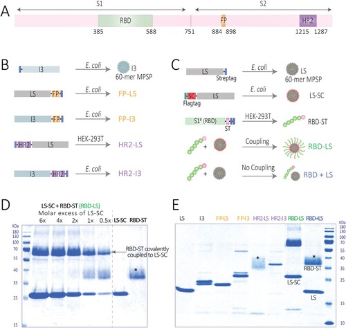
Figure 2. Immunogenicity of MERS-CoV spike MPSP vaccines. (A) Vaccination scheme for rabbit immunizations. Six groups of rabbits (5/group) were vaccinated in a prime/boost regimen with 15 μg of adjuvanted vaccine at 4-week interval and challenged with MERS-CoV (EMC strain; accession no. NC_019843) 3 weeks post-boost. Anti-MERS-CoV spike S2 (B) and S1 (D) IgG titres measured by ELISA in rabbits at different time points. Shown is the mean ± s.e.m. antibody titres from five rabbits per group. (C,E) MERS-CoV neutralizing antibody titres measured by a 90% reduction in a plaque reduction neutralization assay (PRNT90). (B-E) Shown is the mean ± s.e.m. of five rabbits per group. (F,G) Vaccine-induced antibodies in nasal swabs of vaccinated rabbits. Anti MERS-CoV S2 (F) and S1 (G) antibody responses in the nasal swabs (tested at a 1:50 dilution) of vaccinated rabbits pre-challenge (three weeks post-boost). The difference in antibody responses between monomeric (RBD + LS) and multimeric (RBD-LS) RBD was tested for statistical significance using a student’s t-test, with asterisks indicating the level of significance. *P ≤ 0.05. Error bars indicate mean ± s.e.m. The dotted lines represent the limits of detection. HR2, heptad repeat 2; FP, fusion peptide; LS, lumazine synthase 60-meric particles; I3, I3-01 60-meric particles; RBD, receptor binding domain; RBD + LS, monomeric uncoupled RBD; RBD-LS, multimeric RBD coupled to LS through covalent SpyTag/SpyCatcher.
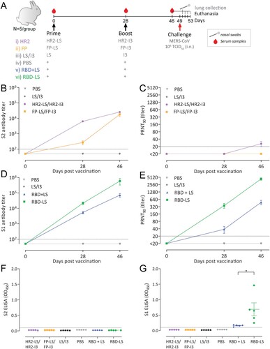
Figure 3. Avidity of vaccine-induced serum antibody responses. The avidity of serum IgG antibody responses after one (A, Day 28) and two immunizations (B, Day 46) with either monomeric RBD (RBD + LS, blue, n = 5) or multimeric RBD (RBD-LS, green, n = 5) was assessed using ammonium thiocyanate (SCN) avidity ELISA. (A, B) The percentage of serum antibodies bound following the addition of different concentration of SCN was used to determine (C) the avidity index (IC50). The difference in serum avidity between both groups was tested for statistical significance using a student’s t-test, with asterisks indicating the level of significance. ***P ≤ 0.001, ****P ≤ 0.0001. Error bars indicate mean ± s.e.m.
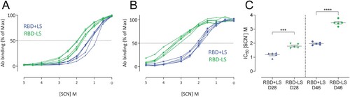
Figure 4. Anti-scaffold antibody responses in sera of vaccinated rabbits. Anti-lumazine synthase (LS) scaffold antibody titres following (A) homologous prime boost in monomeric RBD + LS and multimeric RBD-LS vs heterologous LS/I3 prime boost in control LS/I3 as well as (B) HR2-LS/I3 and FP-LS/I3. Shown are (average ± s.e.m. of n = 5 rabbit/group) antibody titres 4 weeks after prime (day 28, D28) and 3 weeks after boost (day 46, D46) as measured by ELISA. (C) Fold increase (from prime, day 28) in anti-LS antibody titres following boost vaccination (day 46). A paired t-test was performed to determine significant increases in antibody titres post-prime and post-boost within groups (A, B), and an unpaired t-test was performed to determine significant changes in titres between groups (C), with asterisks indicating the level of significance.*P ≤ 0.05, **P ≤ 0.01, ***P ≤ 0.001, ****P ≤ 0.0001. The dotted lines represent the limits of detection. HR2, heptad repeat 2; FP, fusion peptide; LS, lumazine synthase 60-meric particles; I3, I3-01 60-meric particles; RBD, receptor binding domain; RBD + LS, monomeric uncoupled RBD; RBD-LS, multimeric RBD coupled to LS through covalent SpyTag/SpyCatcher.
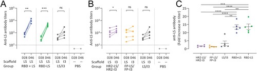
Figure 5. Protective capacity of MERS-CoV MPSP vaccines against upper respiratory tract infection in rabbits. Six groups of vaccinated and control rabbits (n = 5/group) were tested for the presence of viral RNA (A, C) and infectious virus particles (B, D) in the upper respiratory tract (nasal swabs) at days −3 and 1–4 post intranasal viral challenge (days 46 and 50–53 post first vaccination) with 106 TCID50 MERS-CoV EMC strain. Shown is the average ± s.e.m. of five animals per group. The dotted lines represent the limits of detection. HR2, hepad repeat 2; FP, fusion peptide; LS, lumazine synthase 60-meric particles; I3, I3-01 60-meric particles; RBD, receptor binding domain; RBD + LS, monomeric uncoupled RBD; RBD-LS, multimeric RBD coupled to LS through covalent SpyTag/SpyCatcher.
Figure 6. Protective capacity of MERS-CoV MPSP vaccines against lower respiratory tract infection in rabbits. Six groups of vaccinated and control rabbits (n = 5/group) were tested for the presence of viral RNA (A, B) in the lung tissue homogenates and viral nucleocapsid antigen in lung tissues (C) collected 4 days post intranasal viral challenge with 106 TCID50 of MERS-CoV (EMC isolate). (A, B) Shown are the average and SEM equivalent virus titres/gram of tissue. C) Representative pictures of immunohistochemical detection of MERS-CoV nucleoprotein (shown in red) in the lungs of PBS (left) vs RBD-LS (right) immunized rabbits four days post-viral challenge; the upper and lower panels show a 200X and 1000X magnification, respectively. HR2, hepad repeat 2; FP, fusion peptide; LS, lumazine synthase 60-meric particles; I3, I3-01 60-meric particles; RBD, receptor binding domain; RBD + LS, monomeric uncoupled RBD; RBD-LS, multimeric RBD coupled to LS through covalent SpyTag/SpyCatcher.
Supplemental Material
Download ()Data availability
All data are available within the article and its supplementary information or available from the authors on request.