Figures & data
Table 1. Characteristics of Candida auris and Candida albicans isolates used in the study. Bold indicates the isolates used in the fungal burden experiments.
Figure 1. Survival (left) and fungal tissue burden (right) of neutropenic BALB/c mice infected with the four Candida auris clades. In survival studies mice were infected with 19 isolates (South Asian n = 5, East Asian n = 4, South African n = 5 and South American n = 5) representing the four clades. In the graph representing the East Asian clade the “Type strain” represents NCPF 13029 (CBS 10913) type strain isolated from external ear. Fungal kidney, spleen, liver and heart burdens were determined with isolates 196 and 164 from the South Asian, isolates 15 (NCPF 8984) and 12373 (CBS 12373) from the East Asian, isolates 206 (NCPF 13042) and 204 from the South African and isolates I-24 and 16565 (CDC B-16565) from the South American clades at days 2 and 6. The bars represent the medians.
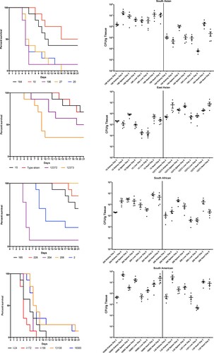
Figure 2. Survival (left) of neutropenic BALB/c mice infected with Candida albicans isolates 3666 and 2606. Fungal kidney, spleen, liver and heart burdens (right) was determined with isolate 3666 at days 2 and 6. The bars represent the medians.
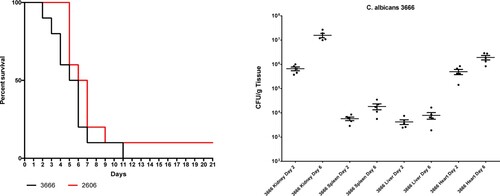
Figure 3. Histopathological examination of the heart, kidney, liver and spleen using Periodic Acid Schiff staining from neutropenic mice intravenously challenged with Candida albicans isolate 3666 (left panels) and C. auris isolate I-24 (right panels), respectively. Histopathological examination was performed six days post infection. In case of C. albicans in the heart, in the kidney and in the spleen blastoconidia and budding yeast cells, pseudohyphae and hyphae were seen. In the liver hyphae were not detected. In the heart both endo- and myocardial involvement is noticeable, the subendocardial myocardium is affected most markedly. Signs of beginning blood vessel invasion in the myocardium is detectable (black arrow). In the kidney C. albicans was detected within the parenchyma, tubuli and glomeruli. In the spleen pseudohyphae and hyphae were seen in the red pulp. C. auris produced large aggregates in the heart, kidney and liver with numerous blastoconidia and budding yeast cells were detected. In the heart coagulative necrosis of myocytes was noticed. In the kidney C. auris cells were detected in the parenchyma, tubuli but not in the glomeruli. In the liver dilated liver sinusoids were filled with yeast cells with central necrosis of lobuli and vacuolar degeneration of hepatocytes. In the spleen yeast cells were seen at the border of the red and white pulp with focal destruction of the white pulp. Small fungal lesions were detectable under the capsule of the spleen and in the sinuses. Magnification × 100.
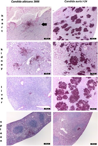
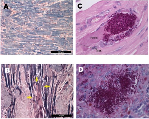
Figure 4. Histological findings from neutropenic mice intravenously challenged with Candida auris. Normal (A) and damaged heart (B) with Mallory’s PTAH staining. PTAH staining performed on the heart of a moribund dissected mouse on day 5 infected with isolate 196 (B) revealed contraction band necrosis or myofibrillar degeneration (yellow arrows). Morphology of C. auris with Periodic Acid Schiff (PAS) staining in the heart’s arterioles on day 1 post infection (C), and in the spleen (D). PAS staining showed blastoconidia and budding yeast cells but never pseudohyphae and hyphae (C–D). Magnification, A-B × 400, C × 1000, D × 400, RBC; red blood cells.