Figures & data
Table 1. Profile of causative microorganisms cultured from postoperative endophthalmitis.
Table 2. Antibiotic susceptibility results of Enterococcus faecalis, Staphylococcus epidermidis, and other coagulase-negative staphylococci (CNS) cultured from postoperative endophthalmitis samples.
Table 3. Profile of conjunctival microbes. Matrix-Assisted Laser Desorption Ionization-Time of Flight Mass Spectrometry (MALDI-TOF MS, Bruker Daltonics GmbH, Germany) was used for identification of microorganisms by identification card (VITEK 2 ID Card, bioMérieux, USA).
Table 4. Antibiotic susceptibility test of S. epidermidis and E. faecalis isolated from human normal conjunctiva to ciprofloxacin, levofloxacin, and moxifloxacin.
Figure 1. Survival assay of S. epidermidis and E. faecalis in in vitro co-culture under various concentrations of moxifloxacin (A). Representative bacterial culture plate images showing the distribution of the bacterial colonies on 0.25 μg/mL moxifloxacin (B) and 16 μg/mL moxifloxacin (C). Colour detection culture plates were used; E. faecalis are shown as blue colonies and S. epidermidis are shown as white colonies. CFU, colony forming unit; MFX, moxifloxacin.
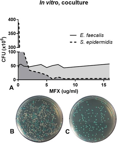
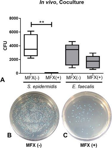
Figure 2. Survival assay of S. epidermidis and E. faecalis in in vivo co-culture after administration of 0.5% moxifloxacin (A). Representative bacterial culture plate images showing the distribution of the bacterial colonies on the absence (B) and presence (C) of moxifloxacin topical drops. Colour detection culture plates were used. E. faecalis are shown as blue colonies and S. epidermidis are shown as white colonies. **, p < 0.005, CFU, colony forming unit; MFX, moxifloxacin; MFX (+), moxifloxacin-treated; MFX (−), moxifloxacin-untreated.