3,085
Views
7
CrossRef citations to date
0
Altmetric
Research Article

Hepatic phaeohyphomycosis due to a novel dematiaceous fungus, Pleurostoma hongkongense sp. nov., and importance of antifungal susceptibility testing

ORCID Icon, ORCID Icon, ORCID Icon, ORCID Icon, ORCID Icon, , ORCID Icon, ORCID Icon & ORCID Icon show all
Pages 81-96 | Received 18 Jun 2020, Accepted 16 Dec 2020, Published online: 17 Jan 2021

Figures & data

Figure 1. Computed tomography image of the upper abdomen. A small fluid collection with gas pocket was found and rim enhancement was observed around the tubal drains.

Figure 1. Computed tomography image of the upper abdomen. A small fluid collection with gas pocket was found and rim enhancement was observed around the tubal drains.

Figure 2. Photomicrographs of the biopsied liver abscess wall tissue. (a) Abundant fungal hyphae, a small amount of necrotic tissue, brown bile pigments as well as a few acute inflammatory cells were observed (Periodic acid-Schiff staining, original magnification 200×). (b) The fungal hyphae were highly septate (Grocott methenamine-silver staining, original magnification 400×).

Figure 2. Photomicrographs of the biopsied liver abscess wall tissue. (a) Abundant fungal hyphae, a small amount of necrotic tissue, brown bile pigments as well as a few acute inflammatory cells were observed (Periodic acid-Schiff staining, original magnification 200×). (b) The fungal hyphae were highly septate (Grocott methenamine-silver staining, original magnification 400×).

Figure 3. Growth of Pleurostoma hongkongense HKU44T on various culture media after 7 days of incubation at 25°C. (a, d) Malt extract agar (MEA). (b, e) Sabouraud dextrose agar (SDA). (c, f) Potato dextrose agar (PDA). Brightness was adjusted individually for each panel.

Figure 3. Growth of Pleurostoma hongkongense HKU44T on various culture media after 7 days of incubation at 25°C. (a, d) Malt extract agar (MEA). (b, e) Sabouraud dextrose agar (SDA). (c, f) Potato dextrose agar (PDA). Brightness was adjusted individually for each panel.

Figure 4. Microscopic features of Pleurostoma hongkongense HKU44T. Arrows indicate notable structures. (a–c) Scanning electron microscopy photographs of notable phialide and conidia features. Scale bars = 5 μm. (d–i) Bright-field microscopy of corresponding features using a slide culture preparation. Slides were prepared via wet mount and stained with lactophenol cotton blue (original magnification 1000×). Scale bars = 10 μm.

Figure 4. Microscopic features of Pleurostoma hongkongense HKU44T. Arrows indicate notable structures. (a–c) Scanning electron microscopy photographs of notable phialide and conidia features. Scale bars = 5 μm. (d–i) Bright-field microscopy of corresponding features using a slide culture preparation. Slides were prepared via wet mount and stained with lactophenol cotton blue (original magnification 1000×). Scale bars = 10 μm.

Figure 5. Phylogenetic tree showing the relationship of Pleurostoma hongkongense HKU44T to its closely related species within the genus Pleurostoma. The tree was inferred from the concatenated sequence data of the internal transcribed spacer (ITS) region, partial β-tubulin gene, partial 18S nrDNA and partial 28S nrDNA by the maximum likelihood method with the substitution model T92 (Tamura 3-parameter model) + G (gamma-distributed rate variation) + I (estimated proportion of invariable sites). The scale bar indicates the estimated number of substitutions per base. Only nodes that were well supported by the maximum-likelihood method (≥70% bootstrap support) have their bootstrap support, calculated from 1,000 replicates, shown; and all these nodes were also well supported by the Bayesian inference method (posterior probabilities ≥0.99). The DDBJ/ENA/GenBank nucleotide accession numbers for Jattaea algeriensis STE-U 6201T are EU367446 (ITS), EU367466 (β-tubulin), EU367462 (18S nrDNA) and EU367456 (28S nrDNA).

Figure 5. Phylogenetic tree showing the relationship of Pleurostoma hongkongense HKU44T to its closely related species within the genus Pleurostoma. The tree was inferred from the concatenated sequence data of the internal transcribed spacer (ITS) region, partial β-tubulin gene, partial 18S nrDNA and partial 28S nrDNA by the maximum likelihood method with the substitution model T92 (Tamura 3-parameter model) + G (gamma-distributed rate variation) + I (estimated proportion of invariable sites). The scale bar indicates the estimated number of substitutions per base. Only nodes that were well supported by the maximum-likelihood method (≥70% bootstrap support) have their bootstrap support, calculated from 1,000 replicates, shown; and all these nodes were also well supported by the Bayesian inference method (posterior probabilities ≥0.99). The DDBJ/ENA/GenBank nucleotide accession numbers for Jattaea algeriensis STE-U 6201T are EU367446 (ITS), EU367466 (β-tubulin), EU367462 (18S nrDNA) and EU367456 (28S nrDNA).

Table 1. Minimum inhibitory concentrations (MICs) or minimum effective concentrations (MECs) of different antifungal agents against Pleurostoma species.Table Footnotea

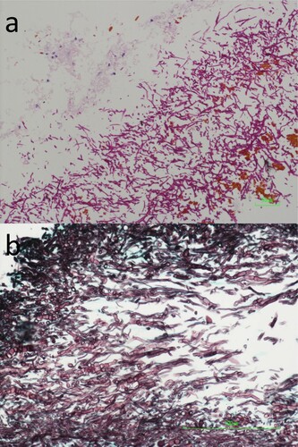
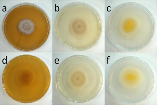
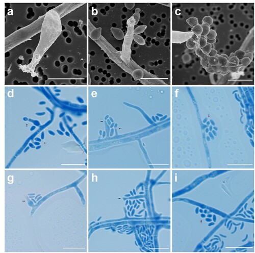
Table 2. Microscopic features of different Pleurostoma species during asexual life cycles.

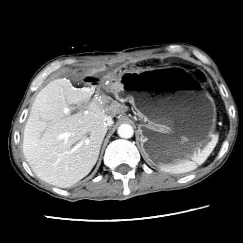
Table 3. Infections caused by Pleurostoma species.

Supplemental material

supplementary_figures.zip

Download Zip (4.5 MB)

Table_S2_revised_final.docx

Download MS Word (14.1 KB)

Table_S1_revised_final.docx

Download MS Word (18.4 KB)