
Figures & data
Figure 1. CL patients exhibit high abundance of components of gamma-secretase complex and inflammatory response genes expression in active lesions. (A) Unbiased RNASeq was performed on skin from 7 Healthy Subjects and lesion from 21 CL patients. Heatmap columns and rows represent each individual and gene, respectively. Heatmap colour reflects z-scores of gene abundance across samples. (B) Gene expression from gamma-secretase complex correlates with the inflammatory response in active lesions. Data from RNASeq (21 CL lesions) was used for correlation matrix between components of gamma-secretase complex and NFKB1, NFKB2, RELA, TNF, IL6, IL1B, IL10, CXCL9, and GZMB genes. Pearson's test was used for correlation statistical analysis and p value is represented according to the size of the circles.; *P < 0.05, **P < 0.01, ***P < 0.001 and ****P < 0.0001.
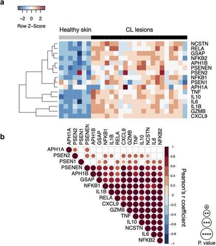
Figure 2. Non-selective gamma-secretase inhibitor (DAPT) decreases inflammatory-associated proteins production from CL patients in response to Leishmania antigens. PBMC from CL patients (n = 12) were cultured in presence or absence of SLA (5 ug/mL) and DAPT (20 µM) for 72 h. The levels of IL-6, IL-1β, TNF, IL-10, CXCL9, and granzyme B were determined in culture supernatants, by ELISA. The black line on the violin plot represents the percentile 50th and the dashed lines, 25th and 75th percentiles, respectively. Statistical analyses were performed using the Wilcoxon test **P < 0.01 and ***P < 0.001.
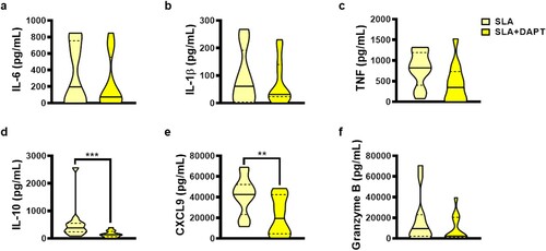
Figure 3. The neutralization of Notch 1 receptor increase production of proinflammatory cytokines from CL patients. PBMC from CL patients (n = 8) were cultured in presence or absence of SLA (5 ug/mL), anti-Notch 1 (20 µg/mL) and anti-Notch 3 (20 µg/mL) for 72 h. The levels of IL-6, IL-1β, TNF, IL-10, CXCL9, and granzyme B were determined in culture supernatants, by ELISA. The black line on the violin plot represents the percentile 50th and the dashed lines, 25th and 75th percentiles, respectively. Statistical analyses were performed using the Wilcoxon test *P < 0.05 and **P < 0.01.
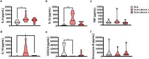
Figure 4. Selective gamma-secretase inhibitor (JLK6) decreases pro-inflammatory cytokine production from CL patients. PBMC from CL patients (n = 15) were cultured in presence or absence of SLA (5 ug/mL) and JLK6 (20 µM) for 72 h. The levels of IL-6, IL-1β, TNF, IL-10, CXCL9, and granzyme B were determined in culture supernatants, by ELISA. The black line on the violin plot represents the percentile 50th and the dashed lines, 25th and 75th percentiles, respectively. Statistical analyses were performed using the Wilcoxon test *P < 0.05, **P < 0.01 and ****P < 0.0001.
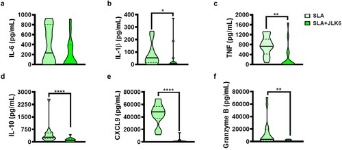
Figure 5. JLK6 downregulates pro-inflammatory cytokines production by lesion cells from CL patients. L. braziliensis lesions skin biopsies from CL patients (n = 5) were cultured in presence or absence of JLK6 (20 uM) for 72 h The levels of IL-6, IL-1β, TNF, IL-10, CXCL9 and granzyme B were determined in culture supernatants, by ELISA. The black line on the violin plot represents the percentile 50th and the dashed lines, 25th and 75th percentiles, respectively. Statistical analyses were performed using the Wilcoxon test *P < 0.05 and **P < 0.01.
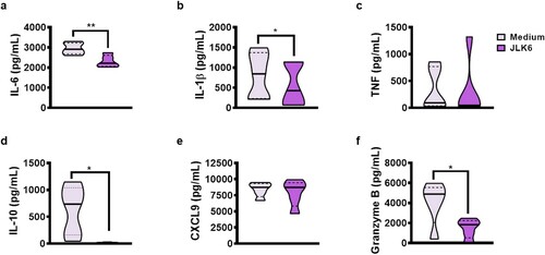
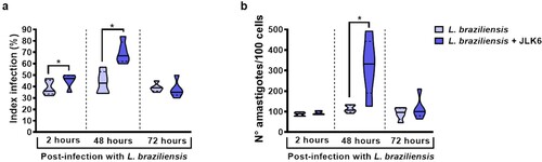
Figure 6. JLK6 does not affect L. braziliensis killing by monocytes from healthy subjects after 72 h. Monocytes from HS (n = 5) were infected with L. braziliensis in stationary phase (ratio 5:1) and cultured in presence or absence of JLK6 (20 µM) for 2, 48 and 72 h. (A) Frequency of infected cells. (B) Number of Leishmania amastigotes/100 monocytes. The black line on the violin plot represents the percentile 50th and the dashed lines represent the 25th and 75th percentiles, respectively. Statistical analyses were performed using the Paired t test *P < 0.05.
Data availability statement
These data is available at https://doi.org/10.6084/m9.figshare.13382723.v1