Figures & data
Figure 1. Phylogenetic tree of whole-genome sequences of BoDV-1 strains. Phylogenetic analysis used the Neighbour-Joining algorithm and p distance model in MEGA 11 [Citation14]. The tree was rooted with the genome sequence of BoDV-2 No/98 (AJ311524). Values at branches represent support in 1000 bootstrap replicates. Only bootstrap values ≥70 at major branches were shown. Names indicated accession number at GenBank, description of isolate, original source, location, and year of isolation. Colour codes indicated designated cluster [Citation11]; 1A = yellow; 2 = green; 3 = pink; 4 = blue. DEU = Germany; JPN = Japan; DEU Federal States: BE = Berlin; BB = Brandenburg; BW = Baden-Wurttemberg; BY = Bavaria; HE = Hesse; NI = Lower Saxony; ST = Saxony-Anhalt.
![Figure 1. Phylogenetic tree of whole-genome sequences of BoDV-1 strains. Phylogenetic analysis used the Neighbour-Joining algorithm and p distance model in MEGA 11 [Citation14]. The tree was rooted with the genome sequence of BoDV-2 No/98 (AJ311524). Values at branches represent support in 1000 bootstrap replicates. Only bootstrap values ≥70 at major branches were shown. Names indicated accession number at GenBank, description of isolate, original source, location, and year of isolation. Colour codes indicated designated cluster [Citation11]; 1A = yellow; 2 = green; 3 = pink; 4 = blue. DEU = Germany; JPN = Japan; DEU Federal States: BE = Berlin; BB = Brandenburg; BW = Baden-Wurttemberg; BY = Bavaria; HE = Hesse; NI = Lower Saxony; ST = Saxony-Anhalt.](/cms/asset/1cfae087-7c79-47eb-afc5-cb214e10e737/temi_a_2065931_f0001_oc.jpg)
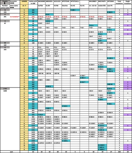
Figure 2. Evaluation of amino acid mutations of BoDV-1 strains. Amino acid (aa) changes of seven human and two laboratory cluster-4 viruses vs. strain V using the one-letter-code. Background colour in turquoise indicates all single changes, in lilac non-conservative single changes. *P indicates a mistake in sequence U04608. P 26 S is the correct reading. S26/S28 is the major phosphorylation site of P-protein. For aa alignments, see Table S2.