
Figures & data
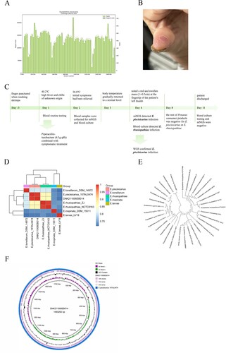
Figure 1. The diagnosis of E. piscisicarius infection and the genome analysis. (A) The whole genome sequencing yielded a total genome coverage of 91.2%. (B) Red and swollen mass on the patient's left thumb fingertip with a size of about 1 × 0.5 cm. (C) The timeline of the clinical course of the patient. (D) ANI heatmap showed that DNK211006EB014 shared an ANI greater than 0.95 with E. piscisicarius_15TAL0474. (E) E. piscisicarius is identified by phylogenetic analysis. (F) Circular map of the genome.
Supplemental material