Figures & data
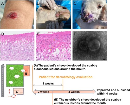
Figure 1. Clinical, histological, TEM findings, and the diagram of the transmission relation chain and course. (A) Physical examination showed central ulceration and a bleeding solitary cutaneous lesion involving the left radiocarpal joint in a diameter of about 1.2 * 0.8 cm with peripheral erythema. (B and C) Similar and typical scabby cutaneous lesions around the mouth in orf virus-infected sheep raised by neighbours were found during further tracking. (D and E) The histological findings displayed partial necrosis, intracellular and intercellular oedema in the epidermis and intraepidermal blistering, diffuse oedema existed in the dermis with dense inflammatory cells infiltration including the neutrophils, lymphocytes and histiocytes into the dermis, also the cytoplasmic eosinophilic inclusion bodies in the epidermis were found. (Haematoxylin-Eosin staining, ×100, ×400). (F) Virus detection by transmission electron microscopy (TEM) analysis at higher magnifications showed typical enveloped virions. Scale bar indicates 50 nm. (G) The three weeks ago when the patient seeking dermatological evaluation, approximately 30 sheep raised by the patient in sheepfold A almost at the same time presented scabby cutaneous lesions around the mouth, also the sheep of neihbours raised in sheepfold B who shared the same pasture also reported an extreme similar history of the sheep presenting cutaneous lesions around the mouth when two weeks after the onset of the lesion in the sheep raised by the patient.