Figures & data
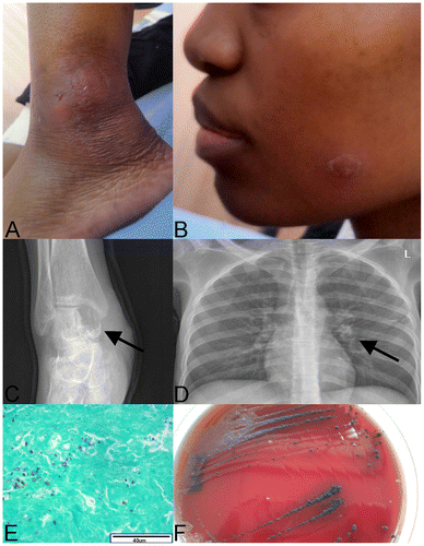
Figure 1: A: Swollen, fluctuant left ankle with sinus formation. B: Well-circumscribed, scaling plaque on the patients face that resolved after treatment with amphotericin B and fluconazole. C: Lytic lesion in the patient’s right talus (arrow), consistent with osteomyelitis. D: Chest X-ray showing a small infiltrate in the left hilar region (arrow). E: Grocott’s stain (40x) showing fibrous connective tissue from the patient’s knee with variable sized fungal elements. F: Aureobasidium pullulans culture on blood agar from blood culture. Colonies were initially small and white, becoming black two weeks later.