Figures & data
Table 1: Summary of epidemiological characteristics of described SSPE cases from published South African studiesCitation 1
Figure 1: Patient was quadrispastic and bedbound.
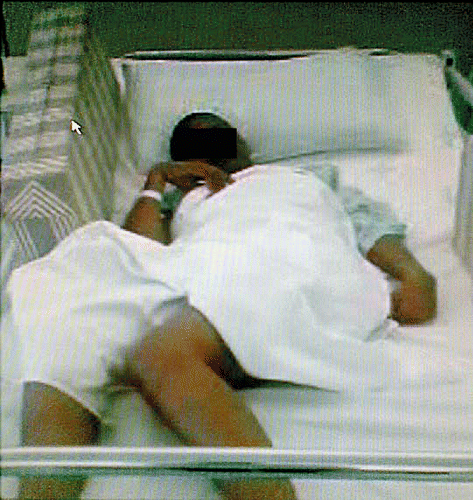
Figure 2: EEG shows a slow background with periodic complexes coincident with patient’s jerks.
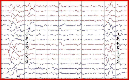
Table 2: Investigations on admission
Figure 3: MRI brain at admission shows periventricular white matter hyperintensities.
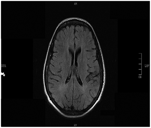
Figure 4: EEG at 9 months post treatment (July 2007) showing significant improvement in the EEG with a normal background without periodic complexes.
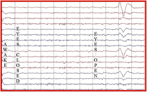
Figure 5: Patient is well, able to walk and converse normally and has no myoclonic jerks.
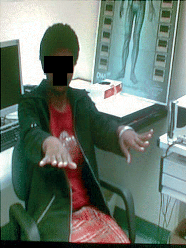
Figure 6: Brain MRI 30 months after treatment (April 2008), showing progressive periventricular white matter hyperintensities.
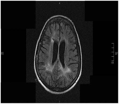
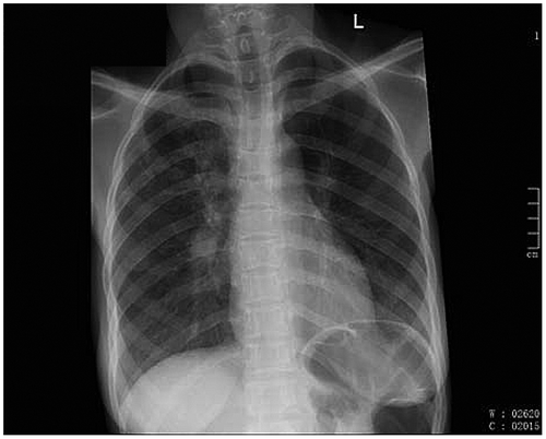
Figure 7: Chest X-ray 30 months after treatment (April 2008), showing hilar infiltrates.