Figures & data
Figure 1. AgNP synthesis using A. herba-alba plant extract, (A) AgNO3 alone, (B) A. herba-alba plant extract and (C) A. herba-alba plant extract with AgNO3. The color of the AgNO3 solution turned from colorless to darkish brown, indicating Ar-AgNP formation.
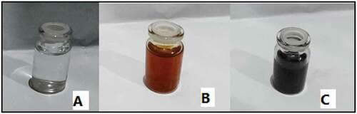
Figure 2. UV-vis absorption spectrum of synthesized AgNPs using A. herba-alba plant extract.
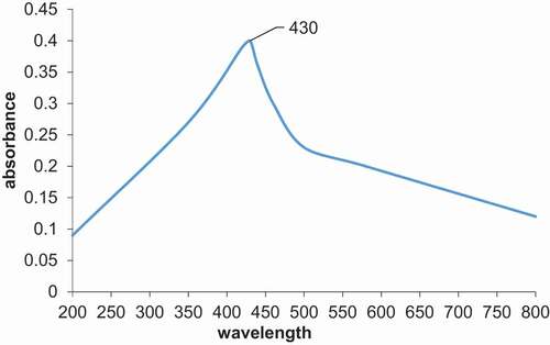
Figure 3. TEM micrographs of synthesized Ar-AgNPs using A. herba-alba plant extract.
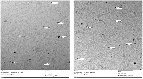
Figure 4. XRD analysis of synthesized Ar-AgNPs using A. herba-alba plant extract.
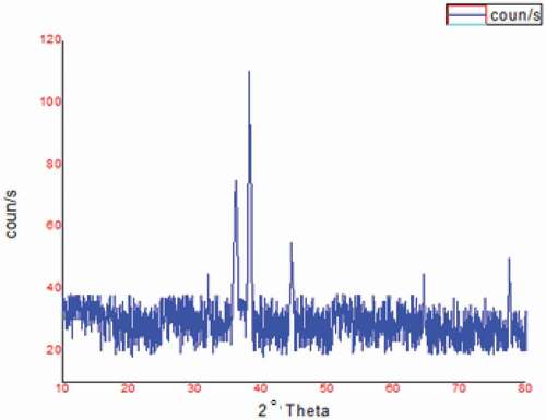
Figure 5. FTIR spectra of A) A. herba-alba plant extract and B) synthesized Ar-AgNPs using A. herba-alba plant extract.
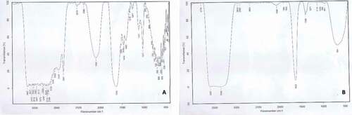
Table 1. Larvicidal activity of the tested solutions against S. littoralis during feeding application
Table 2. Larvicidal activity of the tested solutions against S. littoralis larvae after 30 h using contact application
Table 3. Larvicidal activity of the tested solutions against S. littoralis larvae after 48 h of contact application
Figure 6. Morphological effects of Ar-AgNPs on S. littoralis larvae: (A) control, (B) treatment through feeding and (C) treatment through contact.
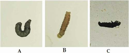
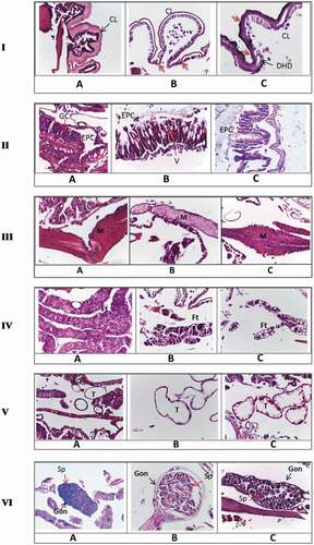
Figure 7. Histopathological changes after treatment S. littoralis larvae with Ar-AgNPs I. (A) Longitudinal section (L.S.) in normal integument showing a normal cuticle layer. (B) L.S. in treated larvae through feeding showing the treated cuticle layer. (C) L.S. in treated larvae through contact showing an irregular cuticle formation, with decolorization of some parts. Red arrows show the destruction of cuticle and hypodermal layers. CL, cuticle layer; HD, hypodermal layer; DHD, destructed hypodermal layer. II. (A) L.S. in normal midgut showing normal gut epithelia (B) T.S. in treated larvae through feeding showing elongation of epithelial cells, nonhomogeneous cytoplasm, and appearance of vacuoles between cells with destructed muscular layer around the gut epithelia. (C) T.S. in treated larvae through contact showing detachment of epithelial cells from the basement membrane, irregular shape of the epithelial cells, and cytoplasmic particles appeared in the lumen. EPC, epithelial cells; GC, goblet cells; v, vacuoles. III. (A) L.S. in normal tissue showing normal muscle tissue (B) L.S. in treated larvae through feeding showing irregular sarcolemma sheath. (C) L.S. in treated larvae through contact showing distortion of muscle. M, muscles.IV. (A) L.S. in normal fat tissue showing normal fat body full of lipid granules (B) L.S. in treated larvae through feeding showing highly affected fat tissue. (C) T.S. in treated larvae through contact showing vacuoles within the fat tissue and the absence of lipid droplets. Ft, fat body. V. (A) L.S. in normal larvae showing normal trachea. (B) L.S. in treated larvae through feeding showing treated trachea. (C) T.S. in treated larvae through contact showing the treated trachea. The cuticle sheath is ruptured and appeared irregular in shape. T, trachea. VI. (A) L.S. in normal gonads. (B) L.S. in treated larvae through feeding showing highly affected gonads. (C) L.S. in treated larvae through contact showing gonads deformation *Gon, gonads; Sp, spermatogonia.