
Figures & data
Figure 1. Illustrate the XRD pattern of silver nanoparticles. GT
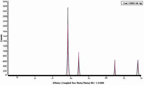
Figure 2. Illustrate the spherical shape of silver nanoparticles with homogenous in shape and size without any agglomeration of silver nanoparticles.
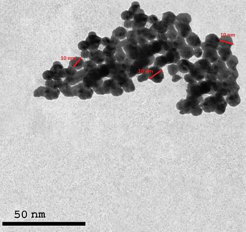
Table 1. Effect of silver nano-particles on weight.
Table 2. Effect of silver nano-particle on hormones.
Table 3. Effect of silver nano-particles on oxidative stress markers in ovarian tissue.
Table 4. Effect of silver nano-particles on oxidative stress markers in uterine tissue.
Figure 3. Photomicrograph of ovarian tissue of control adult female rats showing normal histology of different ovarian sections. GF = Graafian follicle, G = Granulosa, CL = Corpus Luteum, SF = Secondary Follicle, F = Follicle.
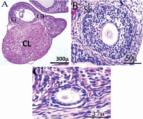
Figure 4. Photomicrograph of ovarian sections of adult female rats treated with (2 mg/kg of AgNps) low dose H&E stain. Showing: from (a to e) Degenerated tertiary follicle (black arrow), the atretic follicle (Orange arrow), the primary follicle (green arrow), high level of vacuolation with degenerated cells (bent arrow), corpus luteum (blue arrow). High percentage of apoptotic cells with pyknotic nuclei (yellow arrow), hemorrhage of blood vessels, dilated blood vessels with many RBCs (white arrow). High vacuolar degeneration in zona granulosa of the secondary follicle and stroma (black arrow), Degeneration in theca interna and cumulus oophorus of the tertiary follicle and slight degeneration in the stroma (black arrow).
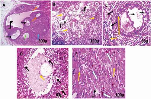
Figure 5. Photomicrograph of ovarian sections of adult female rats treated with (4 mg/kg of AgNps) high dose H&E stain. Showing: from (a to e) corpus luteal (blue), the atretic follicle (Orange arrow), hemorrhage, and dilated blood vessels (green arrow), degenerated stromal cells (double head arrow), Excessive percentage of vacuolation and degeneration (bent arrow), and a high percentage of apoptotic cells with pyknotic nuclei (red arrow). Degenerated tertiary follicle containing antrum (star), degenerated follicle (black arrow).
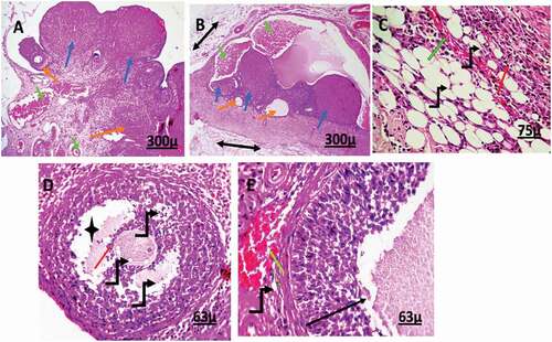
Figure 6. Photomicrograph of uterine tissue of control adult female rats showing normal histology of different uterine sections, normal blood vessels (black arrow), and normal perimetrium layer (head arrow). MI = Myometrium I (longitudinal muscle layer), M II = Myometrium II (circular muscle layer), G = Gland, E = endometrium.
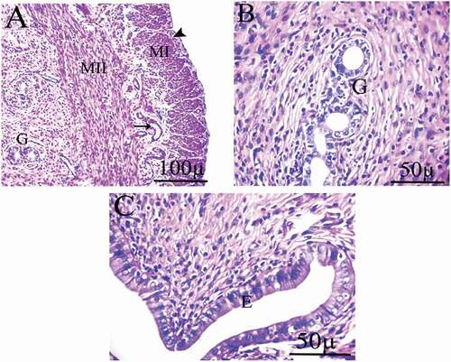
Figure 7. Photomicrographs of uterus of female rats treated with (2 mg/kg AgNps).H&E stain. P = Perimetrium, M = Myometrium, E = Endometrium.
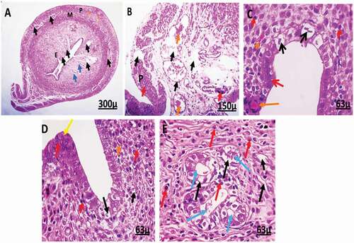
Figure 8. Photomicrographs of uterine tissue of female rats treated with (4 mg/kg AgNps).H&E stain. L = Lumen.
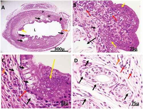
Figure 9. Immunohistochemistry showing caspase-3 streptavidin-biotin- peroxidase staining method in ovarian tissue of control adult female rat showing normal expression of immune stain caspase-3.
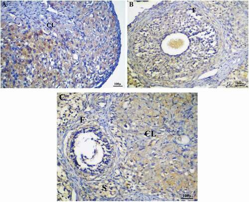
Figure 10. Immunohistochemistry showing caspase-3 streptavidin-biotin- peroxidase staining method in ovarian tissue of adult female rats treated with (2 mg/kg of AgNps) low dose. Showing positive and moderate expression of immune stain caspase-3.
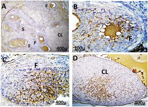
Figure 11. Immunohistochemistry showing caspase-3 streptavidin-biotin- peroxidase staining method in ovarian tissue of adult female rat treated with (4 mg/kg of AgNps) high dose, Showing positive and extreme expression of immune stain caspase-3.
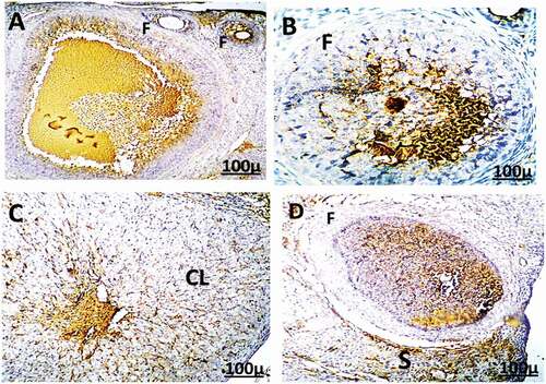
Figure 12. Immunohistochemistry showing caspase-3 streptavidin-biotin- peroxidase staining method in the uterus of control adult female rats showing normal expression of immune stain caspase-3. LM = Longitudinal Muscle, CM = Circular Muscle, E = endometrium, G = Gland.
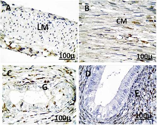
Figure 13. Immunohistochemistry showing caspase-3 streptavidin-biotin- peroxidase staining method in the uterus of adult female rat treated with (2 mg/kg of AgNps) low dose showing positive and moderate expression of immune stain caspase-3.
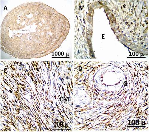
Figure 14. Immunohistochemistry showing caspase-3 streptavidin-biotin- peroxidase staining method in the uterus of adult female rat treated with (4 mg/kg of AgNps) high dose Showing positive and extreme expression of immune stain caspase-3.
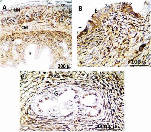
Table 5. Effect of silver nanoparticles on Immunohistochemistry expression for activated caspase-3.