
Figures & data
Figure 1. Transmission electron micrographs of ZnO NPs showing their shape and their size ranged from 7 to 15 nm.
Figure 2. Zeta size of ZnO NPs.
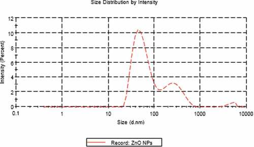
Figure 3. Zeta potential of ZnO NPs.
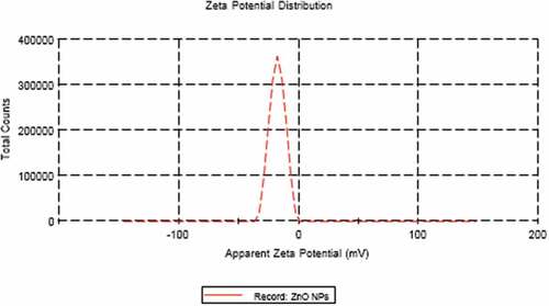
Figure 4. Photomicrographs cross-histological sections of the optic nerve of 21-day-old offspring (a-d) and mother (a1-d1) rats; Control (a&a1) and ZnO NP-treated group (b&b1) showing normal oligodendrocytes and normal astrocyte; 21-day old offspring rats and their mothers treated with LPS showing cytoplasmic vacuolated cells, pyknotic cells, degenerated oligodendrocytes, dilated blood vessels and vacuoles (c&c1); 21-day-old offspring and their mother rats treated with LPS and ZnO NPs showing structural improvement (d&d1). H&E. Abbreviations: AS indicating astrocyte, O indicating oligodendrocyte, V-vacuole; Arrow indicating pyknotic cells; Arrowhead indicating dilated blood vessels; Curved arrow indicating degenerated oligodendrocytes; Zigzag arrow indicating cells with cytoplasmic vacuolation.
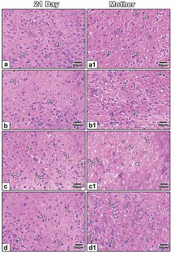
Figure 5. Transmission electron micrographs of the optic nerve of 21-day-old offspring (a-d) and mother (a1-d1) rats; Control (a&a1) and ZnO NPs treated group (b&b1) showing normal nucleus, normal myelinated axons, normal mitochondria; 21-day-old offspring rats their mothers treated with LPS showing degenerated myelinated axons, pyknotic nucleus, dilated rough endoplasmic reticulum and lysosomes (c). Mother treated with LPS showing vacuoles, demyelination, increasing thickness of myelin sheath and pyknotic nucleus (c1). 21-day-old offspring and their mother rats treated with LPS and ZnO NPs showed structural improvement (d&d1). Abbreviations: DMA indicating degenerated myelinated axon; DRER indicating dilated rough endoplasmic reticulum; LY indicating lysosomes; M indicating mitochondria; MA indicating myelinated axon; N indicating nucleus; PN indicating pyknotic nucleus; RER indicating rough endoplasmic reticulum; V indicating vacuoles; Arrow indicating regular nuclear membrane; Arrowhead indicating irregular nuclear membrane; Wavy arrow indicating phagosomes; Tailed arrow indicating thick myelin sheath.
Figure 6. Photomicrographs of formalin-fixed histological sections of the optic nerve immunohistochemical stained with COX-2 of 21-day-old offspring (a-d) and mother (a1-d1) rats. Control (a-a1) and ZnONPs treated group (b-b1) showed a low immunohistochemical reaction of COX-2. 21-day-old offspring and mother rats treated with LPS showed an increased dark brown immunohistochemical reaction of COX-2 (c&c1). 21-day-old offspring and mother rats treated with LPS plus ZnO NPs showed a decreased immunohistochemical reaction of COX-2 (d&d1). Arrowhead indicating the reaction of COX-2.
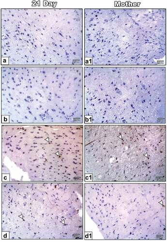
Figure 7. Histogram illustrating the area percent of immunohistochemical reaction of COX-2 in the optic nerve of 21-day-old offspring and their mother rats treated with LPS with or without ZnO NPs. Means with different letters indicate the variations between the groups within the same row using Tukey’s honestly significant difference (p ≤ 0.05) test. Note overexpression of COX-2 in the optic nerve of 21-day-old rats and their mother rats treated with LPS and improved in that treated with ZnO NPs plus LPS. .
Table 1. Biochemical markers of serum of mother rats treated with LPS with or without ZnO NPs.