
Figures & data
Figure 1. a. Brevibacillus borstelensis colonies on agar plat, b. gram positive stain for Brevibacillus borstelensis cells.
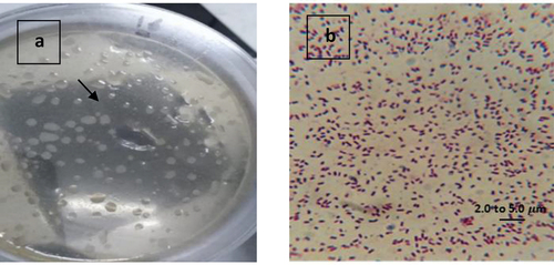
Figure 2. Phylogenetic tree of Brevibacillus borstelensis strain NOB3 using the Neighbor-Joining method.
Table 1. Major features of strains NOB3 and species of the genus Brevibacillus.
Table 2. Antagonistic activity and growth inhibition of Brevibacillus borstelensis strain NOB3.
Figure 3. Effect of Brevibacillus borstelensis NOB3 bioactive compound against different pathogenic microorganisms.
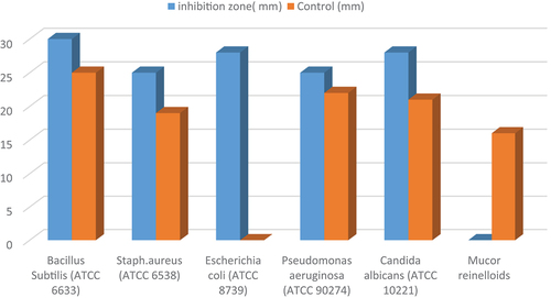
Figure 4. The Minimal Inhibitory Concentration (MIC) of Brevibacillus borstelensis NOB3 bioactive compound.
Figure 5. Effect of bioactive compound of Brevibacillus borstelensis NOB3 on hypotonic solution-induced hemolysis of erythrocyte membrane. P value >0.05.
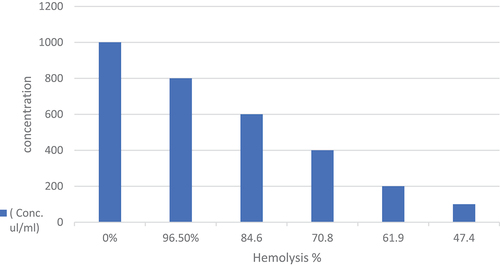
Figure 6. GC-MS chromatogram of Brevibacillus borstelensis NOB3.
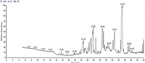
Table 3. GC-MS analysis revealed bioactive compound in an ethyl acetate extract of Brevibacillus borstelensis NOB3.
Figure 7. 3 D of HeLa cell line protein (Cellular tumor antigen p53) retrieved from protein data bank, PDB id: 6VTH, Method: X-RAY DIFFRACTION and Resolution: 2.36 Å.
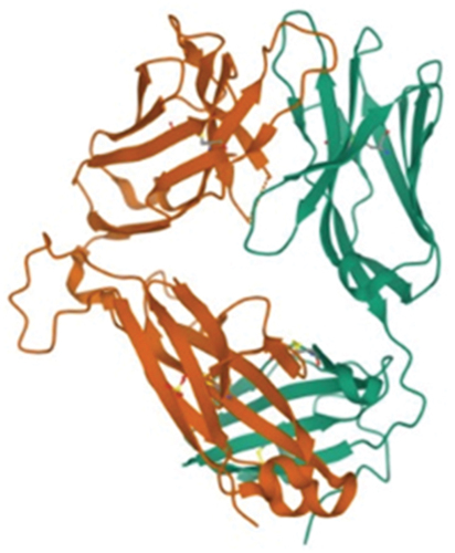
Figure 8. Interaction of the four compounds to the active sites of cervical cancer protein.
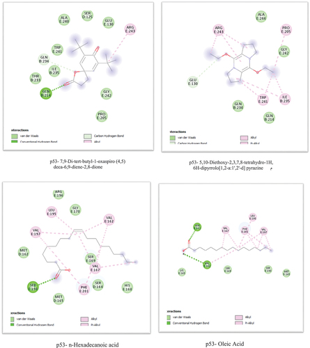
Table 4. In silico docking analysis of bioactive compounds of Brevibacillus borstelensis NOB3against cervical cancer protein.
Figure 9. Concentration of Brevibacillus borstelensis bioactive compounds against Hela cell viability, P value >0.05.
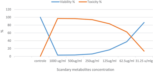
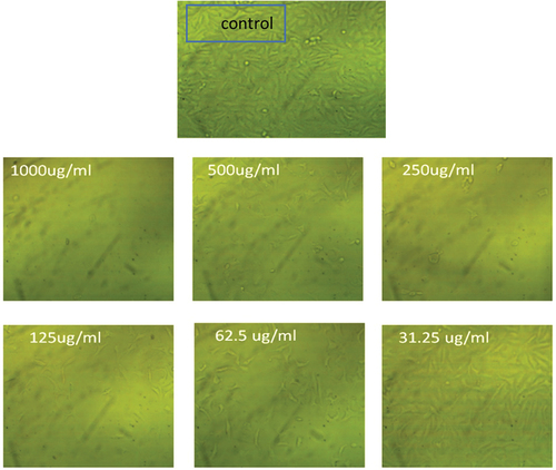
Figure 10. Cell viability and cytotoxicity of Brevibacillus borstelensis NOB3 bioactive compounds on HeLa cell.
Data availability statement
16S rRNA gene sequences supporting the results of this article are available in the GenBank database (https://www.ncbi.nlm.nih.gov/genbank/) under accession numbers ON073840. All other data generated or analyzed during this study are included in this published article.