Figures & data
Figure 1. Electrode locations for the Acticap (red) and EPOC + system (blue), and 4 ROIs chosen for our analyses (Central ROI in orange, left fronto-central ROI in red, right fronto-central ROI in pink, and antero-frontal ROI in purple). Inset in the bottom right shows the double setup on a participant’s head.
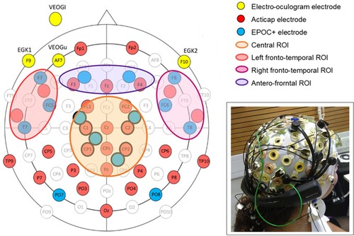
Figure 2. Individual-subject decoding accuracy for a SVM classifier trained to distinguish between matched and mismatched pairs of words-pictures, for Acticap (purple) and EPOC + (yellow) data. Circles indicate the accuracy, with full circles representing significantly above-chance decoding (chance is 50%) according to the permutation test for that participant. Null distributions of accuracy obtained by permutations are shown as a violin plot for each person, with Acticap distribution in light purple and EPOC + distribution in light yellow. Participants are ordered according to Acticap decoding accuracy in this analysis, and this order will remain the same for all other figures.
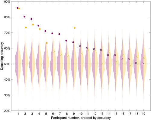
Figure 3. Grand average (N = 19) time-resolved decoding accuracy for a SVM classifier trained to distinguish between matched and mismatched pairs of words-pictures, for Acticap (purple) and EPOC + (yellow) data, shown with standard error of the mean. Time points with significant decoding (p < .05, assessed with threshold-free cluster enhancement permutation tests corrected for multiple comparisons; see Method section) are shown by a purple (Acticap) and yellow (EPOC+) horizontal line at the bottom. Decoding accuracy was significantly above chance for both systems between 200 and 400 ms, extending to 500 ms for Acticap.
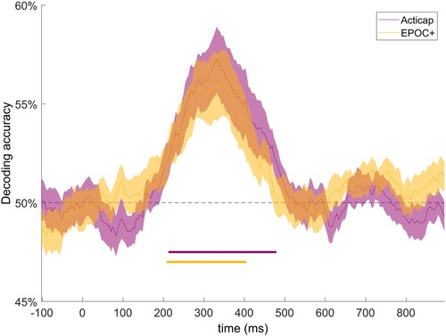
Figure 4. Single-subject time-resolved decoding accuracy for a SVM classifier trained to distinguish between matched and mismatched pairs of words-pictures, for Acticap (purple) and EPOC + (yellow) data. Shaded grey area shows the null distribution of accuracy for Acticap, obtained by permutations. For clarity of presentation, the EPOC + null distribution is not shown, as it looks similar. Time points with significant decoding (p < .05, assessed with threshold-free cluster enhancement permutation tests corrected for multiple comparisons; see Methods) are shown by a purple (Acticap) and yellow (EPOC+) horizontal line at the bottom of each plot. Decoding accuracy was significantly above chance in at least one cluster of time points for 7/19 participants using Acticap, and for 6 participants using EPOC + .
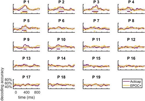
Figure 5. Grand average (N = 19) ERP response to pictures that followed matched (blue) and mismatched (red) words. Results for four regions of interests (ROI) are shown for Acticap (top) and EPOC + (bottom). Time-points with a significant difference between conditions are indicated with an orange horizontal line, after Bonferroni correction for 4 tests (one test per ROI). Group-level N400 effects are present in all 4 ROIs for both EEG systems, with markedly similar timecourses and amplitudes between the two systems.
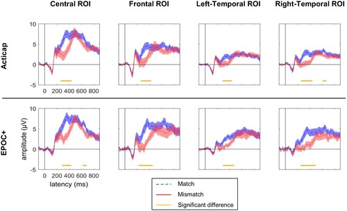
Figure 6. Individual-subject ERP responses to pictures that followed matched (blue) and mismatched (red) words, for a central ROI for Acticap (columns 1 and 3), and EPOC + (columns 2 and 4). Time-points with a significant difference between match and mismatch conditions are shown as a black horizontal line. In this ROI, 8 participants show an N400 effect with Acticap, and 5 participants with EPOC + .
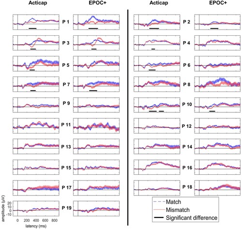
Table 1. Intraclass correlation values and 95% confidence interval (CI) for the ERP recorded in each ROI and each condition across the two EEG systems.
Figure 7. Grand-average topographic distribution of N400 effects across time. Blue colours indicate more negative responses for the mismatched condition, and yellow colours indicate more negative responses for the matched condition. An N400 effect (mismatched < matched) is seen in parietal to frontal central regions, and is strongest from 300 to 500 ms post-stimulus onset.
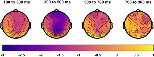
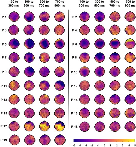
Figure 8. Individual-subject topographic distribution of N400 effects across time. Blue colours indicate more negative responses for the mismatched condition, and yellow colours indicate more negative responses for the matched condition. N400 effect location and timing varied slightly across individuals.