Figures & data
Figure 1. Foc.us tDCS headset.
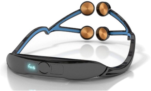
Figure 2. A simple diagram illustrating the basics of tDCS. Two electrodes, cathodal or anodal, are hooked up to a battery and placed on certain regions of the skull. Current is then applied through the electrodes to alter neural activity. Source: George and Aston-Jones (Citation2010).
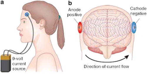
Figure 3. (A) Schematic for Luigi Galvani’s eighteenth-century Galvanic Battery. (B) Details from plate V in Aldini (Citation1804). Sources: A, The Popular Science Monthly, Volume 12, 1877; B, Parent (Citation2004).
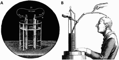
Figure 4. Electrical stimulation of the ventral medial prefrontal cortex caused activation of the ventral midbrain. Source: Chib et al. (Citation2013).
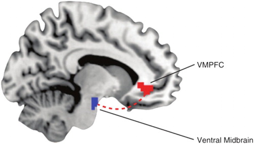
Figure 5. (A) Electrical stimulation of the cortex. (B) Induced dopamine release in the caudate nucleus due to stimulation of the cortex. Source: Strafella et al. (Citation2001).
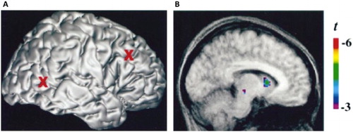
Figure 6. (A) An inhibitory neuron is inhibiting the firing on the excitatory neuron. This is in turn in preventing the firing of another excitatory neuron. (B) Cathodal stimulation is inhibiting the inhibitory neuron (disinhibition), which allows the excitatory neuron to fire and activate another excitatory neuron. Source: Author generated.
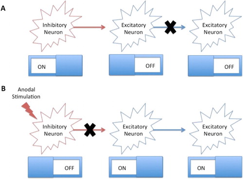
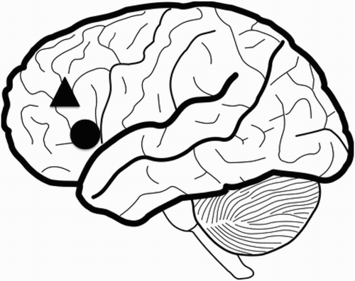
Figure 7. Broca’s area (represented by the circle) is within close proximity of the dorsolateral prefrontal cortex (represented by the triangle). Unintentional stimulation of either area may occur while attempting to use tDCS. Source: Author generated.