768
Views
3
CrossRef citations to date
0
Altmetric
Research Article

Coral-associated bacterial extracts inhibit cellular NF-κB pathway

ORCID Icon, , , & | (Reviewing Editor)
Article: 1292865 | Received 30 Nov 2016, Accepted 06 Feb 2017, Published online: 28 Mar 2017

Figures & data

Figure 1. Bacterial extracts were tested by the NF-kB luciferase reporter gene assay. The results were compared to control, vehicle treated cells (0.02% MeOH).
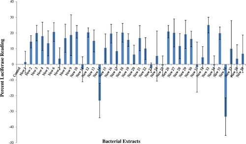
Figure 1. Bacterial extracts were tested by the NF-kB luciferase reporter gene assay. The results were compared to control, vehicle treated cells (0.02% MeOH).

Figure 2. Detection of NF-kB canonical and alternative pathway subunits in L428 cells following New-33 treatment. Western blot analysis of NF-kB subunits from cytoplasm and nuclei of L428 cells treated with 0.02% MeOH or increasing concentrations of the extract was performed. In order to detect NF-κB nuclear translocation, separate nuclear and cytoplasmic lysates (10 × 10 power 6 cells per sample) were prepared using the NucBuster kit (Novagen). Protein concentrations were estimated using the Bradford method (BioRad). Detection of NF-κB canonical subunits in cytoplasm (A) and nuclei (B).
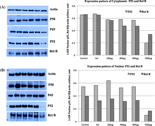
Figure 2. Detection of NF-kB canonical and alternative pathway subunits in L428 cells following New-33 treatment. Western blot analysis of NF-kB subunits from cytoplasm and nuclei of L428 cells treated with 0.02% MeOH or increasing concentrations of the extract was performed. In order to detect NF-κB nuclear translocation, separate nuclear and cytoplasmic lysates (10 × 10 power 6 cells per sample) were prepared using the NucBuster kit (Novagen). Protein concentrations were estimated using the Bradford method (BioRad). Detection of NF-κB canonical subunits in cytoplasm (A) and nuclei (B).

Figure 3. Scratch assay on MCF-7 human breast cancer cell line following New-33 treatment. MCF-7 monolayers were scratched and incubated with ethanol or New-33 extracts at two concentrations. The cells were photographed every 24 h to observe gap closure.
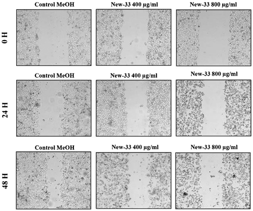
Figure 3. Scratch assay on MCF-7 human breast cancer cell line following New-33 treatment. MCF-7 monolayers were scratched and incubated with ethanol or New-33 extracts at two concentrations. The cells were photographed every 24 h to observe gap closure.
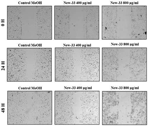
Figure 4. Scratch assay on A549 human lung carcinoma cell line following New-33 treatment. A549 monolayers were scratched and incubated with ethanol or New-33 extracts at two concentrations. The cells were photographed every 24 h to observe gap closure.
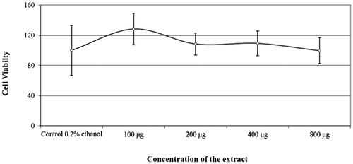
Figure 4. Scratch assay on A549 human lung carcinoma cell line following New-33 treatment. A549 monolayers were scratched and incubated with ethanol or New-33 extracts at two concentrations. The cells were photographed every 24 h to observe gap closure.
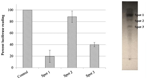
Figure 5. Effect of New-33 extract on the cell viability of L428 cells at 24 hours.

Figure 5. Effect of New-33 extract on the cell viability of L428 cells at 24 hours.

Figure 6. Thin layer chromatographic separation of New-33 extract and the TLC purified fractions visualized under UV were extracted individually using acetonitrile and were assayed for NF-κB modulatory capability using NF-κB luciferase reporter gene assay.

Figure 6. Thin layer chromatographic separation of New-33 extract and the TLC purified fractions visualized under UV were extracted individually using acetonitrile and were assayed for NF-κB modulatory capability using NF-κB luciferase reporter gene assay.