57,981
Views
28
CrossRef citations to date
0
Altmetric
Research Article

Brain changes during a shamanic trance: Altered modes of consciousness, hemispheric laterality, and systemic psychobiology

, & | (Reviewing Editor)
Article: 1313522 | Received 30 Dec 2016, Accepted 24 Mar 2017, Published online: 24 Apr 2017

Figures & data

Figure 1. Ordinary (OSC) vs. altered (ASC) states of consciousness as a function of arousal/absorption.
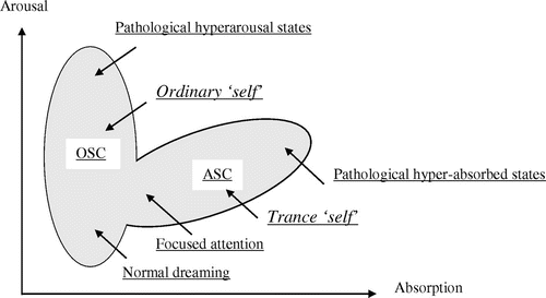
Figure 1. Ordinary (OSC) vs. altered (ASC) states of consciousness as a function of arousal/absorption.

Table 1. EEG frequency bands and sub-bands as defined in this study

Figure 2. Centroid analysis. (A) Centroid analysis of the subject in the trance state (red) vs. 80 normal dextral female controls (blue). (B) Discriminant score comparison of the subject in the trance state (red) vs. 80 normal dextral female controls (blue) and 84 females with depression (yellow). (C) Discriminant score comparison of the subject in the trance state (red) vs. 80 normal dextral female controls (blue) and 19 females with mania (yellow). (D) Discriminant score comparison of the subject in the trance state (red) vs. 80 normal dextral female controls (blue) and 18 females with schizophrenia (yellow).
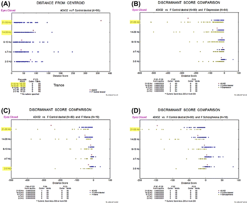
Figure 2. Centroid analysis. (A) Centroid analysis of the subject in the trance state (red) vs. 80 normal dextral female controls (blue). (B) Discriminant score comparison of the subject in the trance state (red) vs. 80 normal dextral female controls (blue) and 84 females with depression (yellow). (C) Discriminant score comparison of the subject in the trance state (red) vs. 80 normal dextral female controls (blue) and 19 females with mania (yellow). (D) Discriminant score comparison of the subject in the trance state (red) vs. 80 normal dextral female controls (blue) and 18 females with schizophrenia (yellow).

Figure 3. Quantitative EEG mapping of the subject in the trance state (left 3 × 3 maps) vs. 78 normal dextral female controls (right 3 × 3 maps): (A) Low beta band, EC condition. (B) High beta band, EC condition. (C) The subject in the trance state (left 3 × 3 maps) vs. in her normal state (right 3 × 3 maps): high beta band, EC condition.
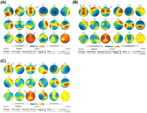
Figure 3. Quantitative EEG mapping of the subject in the trance state (left 3 × 3 maps) vs. 78 normal dextral female controls (right 3 × 3 maps): (A) Low beta band, EC condition. (B) High beta band, EC condition. (C) The subject in the trance state (left 3 × 3 maps) vs. in her normal state (right 3 × 3 maps): high beta band, EC condition.

Figure 4. LORETA power source distribution of the subject in the normal state vs. 78 normal dextral female controls, EC condition: (A) Delta band; (B) Theta band; (C) Alpha band; (D) Low beta band; (E) High beta band; (F) Gamma band.
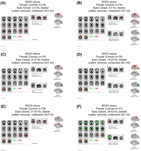
Figure 4. LORETA power source distribution of the subject in the normal state vs. 78 normal dextral female controls, EC condition: (A) Delta band; (B) Theta band; (C) Alpha band; (D) Low beta band; (E) High beta band; (F) Gamma band.
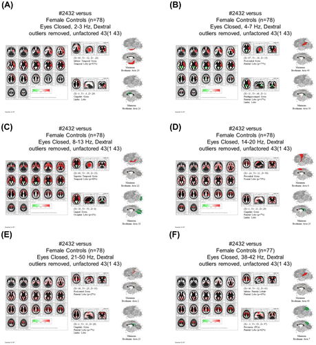
Figure 5. LORETA power source distribution of the subject in the trance state vs. 78 normal dextral female controls, EC condition: (A) Delta band; (B) Theta band; (C) Alpha band; (D) Low beta band; (E) High beta band; (F) Gamma band.
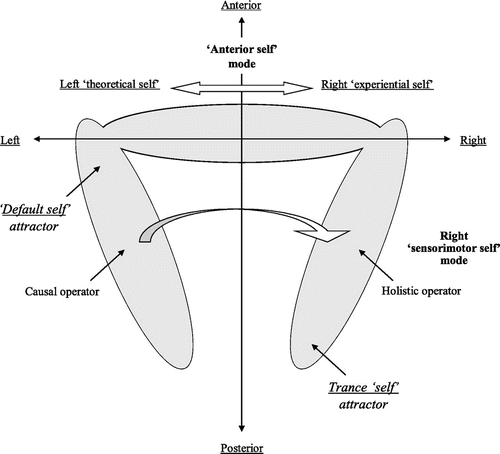
Figure 5. LORETA power source distribution of the subject in the trance state vs. 78 normal dextral female controls, EC condition: (A) Delta band; (B) Theta band; (C) Alpha band; (D) Low beta band; (E) High beta band; (F) Gamma band.

Figure 6. Posterior–anterior and lateral axes in normal and altered states of consciousness.

Figure 6. Posterior–anterior and lateral axes in normal and altered states of consciousness.

Table 2. Lateralized log R/L alpha power ratios in normal vs. trance state