Figures & data
Figure 1. X-ray diffraction spectra of Zn0.7MnxNi0.3−xO (x = 0.05, 0.1, 0.15, 0.2) nanoparticles calcined at 700°C.
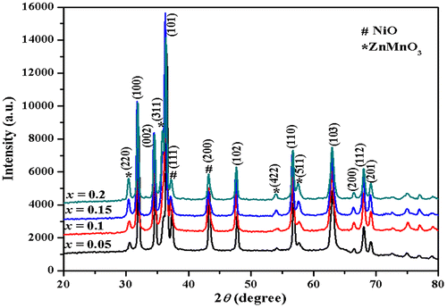
Table 1. Compositional dependence of average crystalline size (D), SEM (D), optical band gap (Eopt), activation energy of grain boundaries (Egb), grains (Eg), coercive field (Hc), remanent magnetization (Mr), and saturation magnetization (Ms)
Figure 2. Scanning electron micrograph (SEM) of Zn0.7MnxNi0.3−xO (x = 0.1) sample.
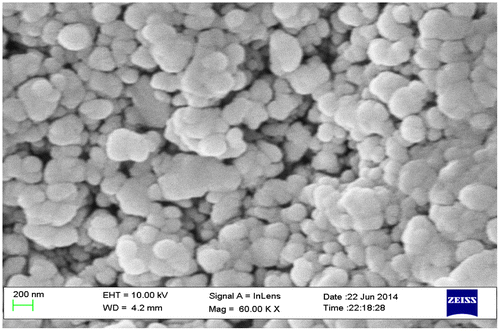
Figure 3. (F(R)hυ)2 vs. hυ plot for direct band gap determination of Zn0.7MnxNi0.3−xO (x = 0.05, 0.1, 0.15, 0.2) nanoparticles.
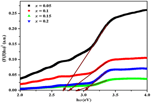
Figure 4. Variation of AC conductivity for Zn0.7MnxNi0.3−xO (Pure ZnO, x = 0.0, 0.05, 0.1, 0.15, 0.2) samples at 323 K.
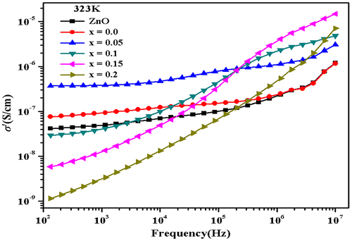
Figure 5. Measured total AC conductivity (σ′) for Zn0.7Mn0.05Ni0.25O composition, shown as function of frequency at eight different temperatures.
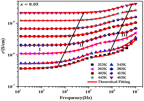
Figure 6. Plot of frequency exponent n with temperature for Zn0.7MnxNi0.3−xO (x = 0.05, 0.1, 0.15, 0.2) samples at low frequency dispersion region.
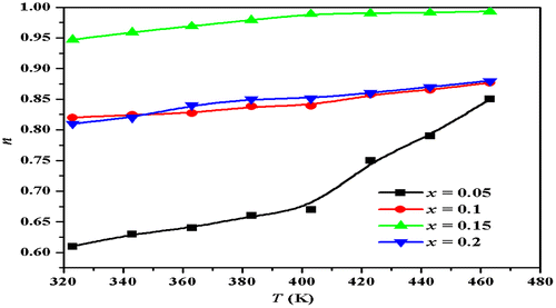
Figure 7. Arrhenius plots of DC conductivity (σdc) for Zn0.7MnxNi0.3−xO (x = 0.05, 0.1, 0.15, 0.2) samples.
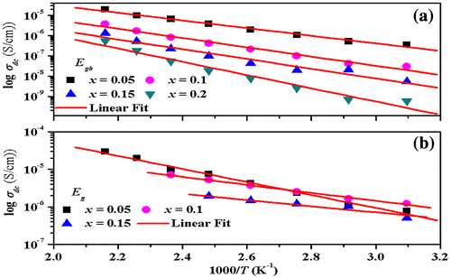
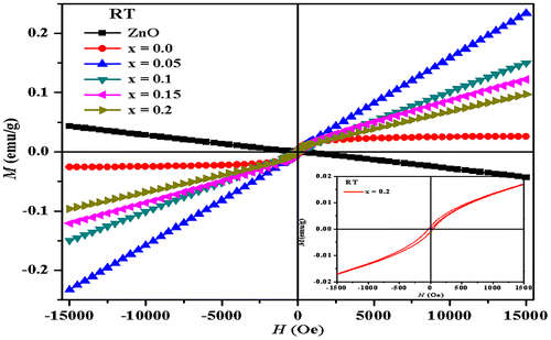
Figure 8. Field-dependent magnetization at room temperature Zn0.7MnxNi0.3−xO (Pure ZnO, x = 0.0, 0.05, 0.1, 0.15, 0.2) samples.