Figures & data
Figure 1. FESEM images of CNTs synthesised at precursor vaporisation temperatures of (a) 370, (b) 420, (c)–(d) 470, (e)–(f) 520, (g)–(h) 570, (i)–(j) 670 and (k)–(l) 770°C at different magnification.
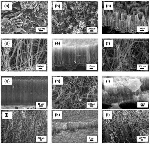
Figure 2. Micro-Raman spectra of the samples synthesised at different vaporisation temperatures of 370–770°C with enlarged spectrum of 2D peak.
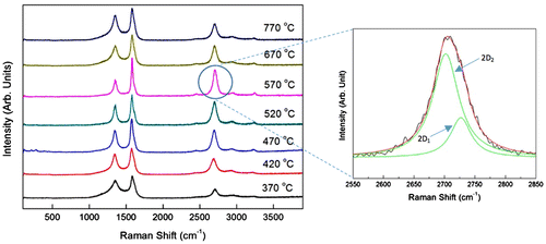
Table 1. The ID/IG ratio of the CNTs samples synthesised at different vaporisation temperatures of 370–770°C
Table 2. The curve fitting results for second-order Raman peak using the Lorentzian distribution function
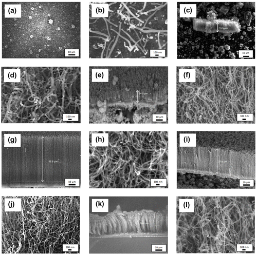
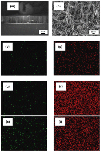
Table 3. Diameter, length and the growth rate of the CNTs synthesised using different catalyst concentration of 1.33–10.33 wt%
Figure 4. Micro-Raman spectra of the sample synthesised using different catalyst concentration of 1.33–10.33 wt%.
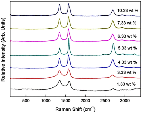
Table 4. The D and G peaks position and the ID/IG ratio of the sample synthesised using catalyst concentration of 1.33–10.33 wt%.
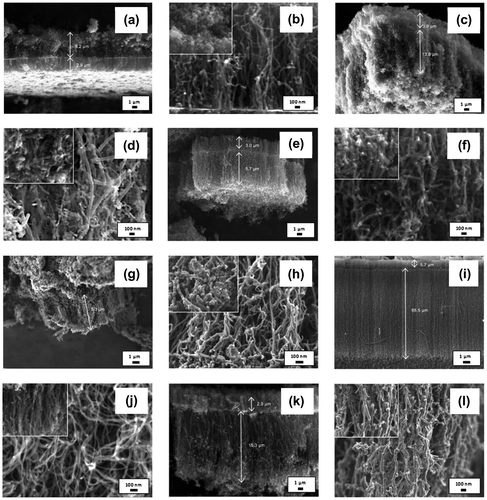
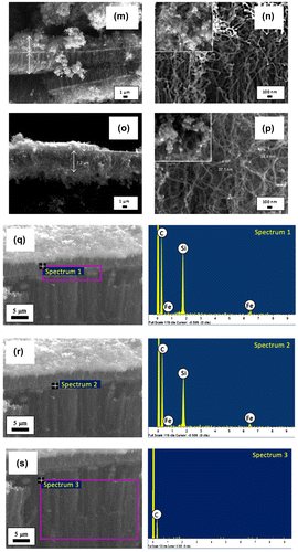
Table 5. The weight % of element content at different part of VACNTs
Table 6. Diameter, length and the growth rate of the nanotubes synthesised by synthesis time of 5–90 min
Figure 6. Micro-Raman spectra of the samples synthesised at various synthesis time of 5–90 min.
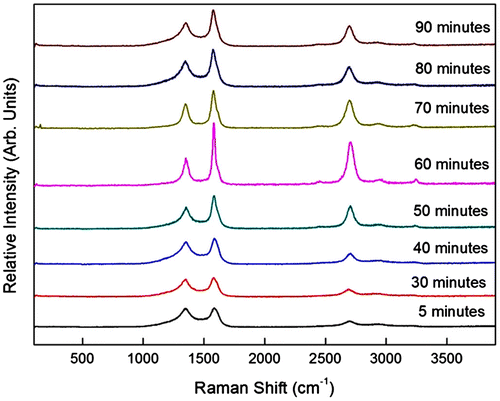
Table 7. The D, G and G′ peaks width and the ID/IG ratio of the sample synthesised by synthesis time of 5–90 min
Figure 7. TGA and DTGA curves of VACNTs synthesised using optimum parameters.
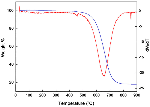
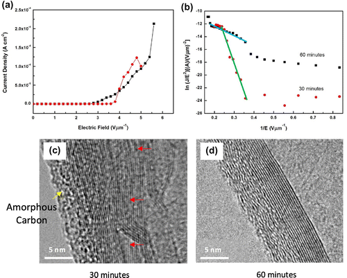
Figure 8. (a) Typical J-E curves, (b) F-N plot and HR-TEM images of the VACNTs from waste chicken fats by (c) 30 and (d) 60 min.