Figures & data
Figure 1. Chemical structures of the drugs.
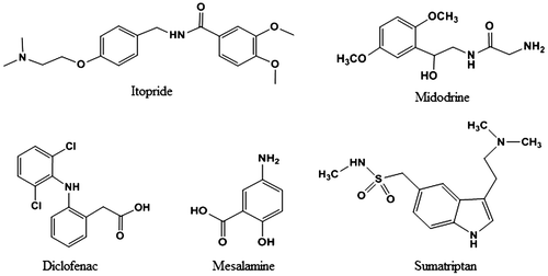
Figure 2. Representative absorption spectra of studied drugs (ITO (20 µg/mL), MID (120 µg/mL), DIC (20 µg/mL), MES (30 µg/mL), and SUM (100 µg/mL)) and BTB (0.1%) in aqueous medium.
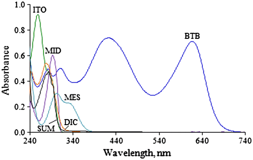
Figure 3. Ion-pair complex formation of BTB with (a) ITO (3.0–30 µg/mL), (b) MID (1.0–20 µg/mL), (c) DIC (1.5–40 µg/mL), (d) MES (1.2–12 µg/mL), and (e) SUM (0.5–15 µg/mL)] in chloroform.
![Figure 3. Ion-pair complex formation of BTB with (a) ITO (3.0–30 µg/mL), (b) MID (1.0–20 µg/mL), (c) DIC (1.5–40 µg/mL), (d) MES (1.2–12 µg/mL), and (e) SUM (0.5–15 µg/mL)] in chloroform.](/cms/asset/583283ae-7dcf-44ae-b9bb-cf162bb50f40/oach_a_1075852_f0003_oc.gif)
Figure 4. Plausible mechanism of ion-pair complex formation between the drugs and the anionic dye, bromothymol blue.
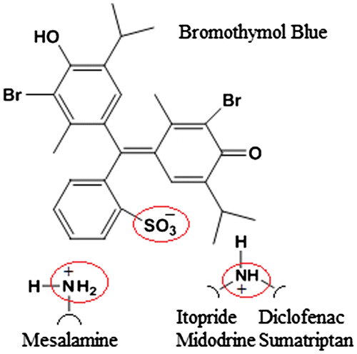
Figure 5. Job’s plot for ion-pair complexes of BTB with (a) ITO, (b) MID, (c) DIC, (d) MES and (e) SUM in chloroform (1.5 × 10⁻³ M).
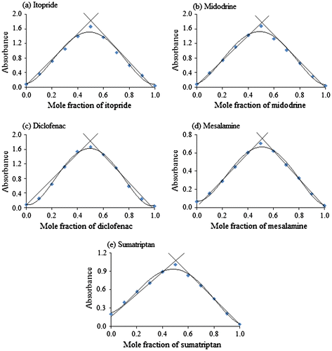
Figure 6. Benesi–Hildebrand plots for association constants of ion-pair complexes between the drugs and BTB.
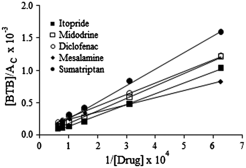
Table 1. Association constants of the ion-pair complexes between the drugs and BTB
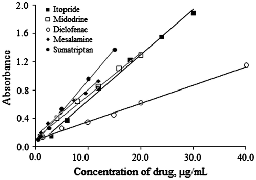
Figure 7. Calibration curves for ITO, MID, DIC, MES, and SUM at 411, 410, 413, 412, and 414 nm, respectively.