Figures & data
Figure 1. SDS-PAGE analysis of the purified lectins. (a) 12% SDS-PAGE analysis of the purified DLL-I; Lane 1: molecular weight marker (kDa), Lane 2: purified lectin. (b) 10% SDS-PAGE analysis of the purified DLL-II; Lane 1: protein molecular weight marker (kDa), Lane 2: purified protein. (c) 10% SDS-PAGE analysis of the purified lactose-specific lectin; Lane 1: molecular weight marker (kDa); Lane 2: purified lectin (*Possible lactose-binding protein). Arrows indicate the subunits of the Unio lectin. (d) 10% SDS-PAGE analysis of the purified WGA lectin; Lane1: purified WGA lectin, Lane 2: molecular weight marker.
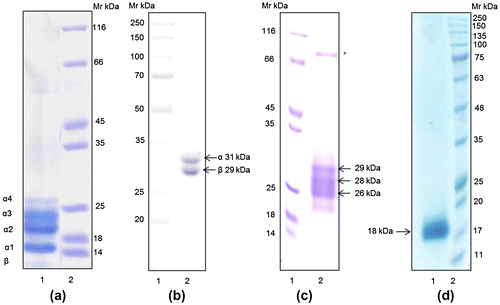
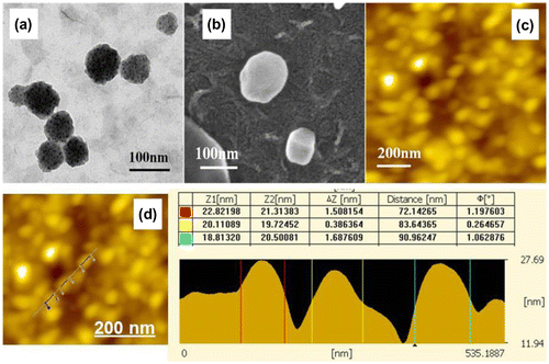
Figure 2. TEM, SEM, and AFM micrographs of the purified DLL-I nanoparticles. (a) Transmission electron micrograph of the purified DLL-I lectin nanoparticle. (b) Scanning electron micrograph of the purified DLL-I lectin nanoparticle. (c) AFM images of DLL-I nanoparticles. (d) Cross-sectional line profile of the AFM image of DLL-I lectin nanoparticles.
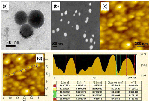
Figure 3. TEM, SEM, and AFM micrographs of the purified DLL-II nanoparticles. (a) Transmission electron micrograph of the purified DLL-II lectin nanoparticles. (b) Scanning electron micrograph of the purified DLL-II lectin nanoparticles. (c) AFM images of DLL-II lectin nanoparticles. (d) Cross-sectional line profile of the AFM image of DLL-II lectin nanoparticle.
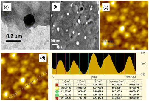
Figure 4. TEM, SEM, AFM, and SNOM micrographs of LSL nanoparticles. (a) & (b) Transmission electron micrographs of the LSL nanoparticles. (c) Scanning electron micrograph of the lactose-specific lectin nanoparticles. (d) AFM image of the lactose-specific lectin nanoparticles. (e) & (f) SNOM images of the lactose-specific lectin nanoparticles.
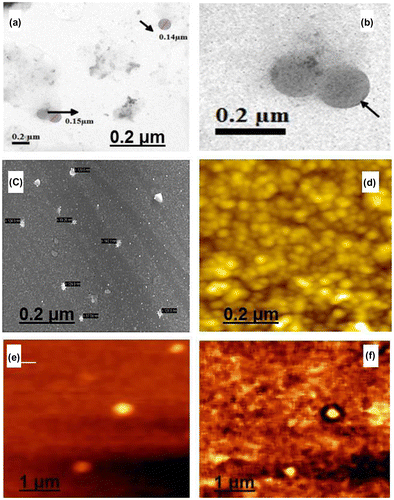
Figure 5. TEM, SEM, and AFM micrographs of the purified WGA lectin nanoparticles. (a) Transmission electron micrograph of the purified WGA lectin nanoparticles. (b) Scanning electron micrograph of the purified WGA lectin nanoparticles. (c) AFM images of WGA lectin nanoparticles. (d) Cross-sectional line profile of the AFM image of WGA lectin nanoparticles.