Figures & data
Figure 1. Stressors activate forebrain and brainstem regions to prepare for “fight-or-flight”.
Notes: Acute injury or a perceived threat leads to activation of psychogenic and/or homeostatic stress. Psychogenic stress recruits areas in the limbic areas of the forebrain, including the hippocampus and the amygdala, which influence the activity of hypothalamic nuclei. A homeostatic challenge activates brainstem nuclei, which either relay this information to hypothalamic regions or directly to sympathetic preganglionic neurons in the spinal cord. The hypothalamus receives modulatory input from forebrain and medullary nuclei and is the main driver of the HPA axis (in blue) and the sympathetic nervous system (SNS, in green). Release of corticotropin-releasing hormone (CRH) from parvocellular neurons in the paraventricu lar nucleus of the hypothalamus on cells expressing adrenocorticotropic hormone (ACTH) leads to release of ACTH into the bloodstream via the blood, ACTH reaches the adrenal cortex where it stimulates the release of corticosterone (cortisol in humans). Corticosterone mobilises glucose by increasing gluconeogenesis and by counteracting the effects of insulin. Parallel activation of the SNS (green) leads to release of adrenalin from postganglionic sympathetic neurons onto target tissues, including blood vessels, the adrenal medulla, heart and the respiratory system. This leads to increased muscle blood flow and decreased blood flow to the organs in the abdominal cavity (splanchnic blood flow) ensuring sufficient oxygen and energy supply to the muscles. The secretion of adrenaline from the adrenal medulla into the blood further increases heart and respiratory rate and the mobilisation of glucose through glycogenolysis and lipolysis.
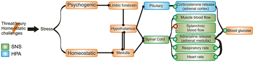
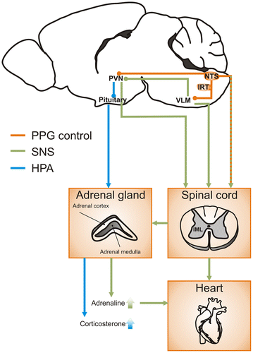
Figure 2. PPG pathways to activate both HPA axis and sympathetic nervous system in the control of stress responses.
Notes: PPG neurons in the nucleus of the solitary tract (NTS) and the intermediate reticular nucleus (IRT) are activated by stressors. Activation of PPG neurons leads to release of GLP-1 (orange) onto parvocellular cells in the PVN, which in turn activate the HPA axis (blue arrows) by stimulating cells in the pituitary to release adrenocorticotropic hormone (ACTH). ACTH acts on the adrenal cortex to increase the secretion of corticosterone. In parallel, PPG neurons send descending axons directly to spinal sympathetic preganglionic neurons in the intermediolateral column (IML) and ascending axons to presympathetic neurons in VLM and PVN. Recruitment of PVN and VLM neurons also leads to activation of the sympathetic nervous system (green arrows) via descending fibres to the sympathetic preganglionic neurons in the IML in the spinal cord, which in turn increase heart rate and stimulate the release of adrenaline from the adrenal medulla.