Figures & data
Table 1. Total phenol content of the Myrothamnus flabellifolius extracts
Figure 1. TLC-DPPH assay showing the anti-oxidant activities of different concentrations of MF spotted on a TLC sheet.
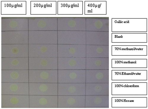
Figure 2. Comparative DPPH scavenging activity of Gallic acid and Myrothamnus flabellifolius.
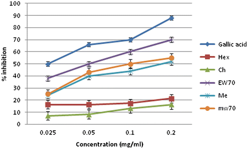
Table 2. In vitro ABTS free radical scavenging activity of various extracts of Myrothamnus flabellifolius
Figure 3. Comparative reducing power of Gallic acid and Myrothamnus flabellifolius leaf.
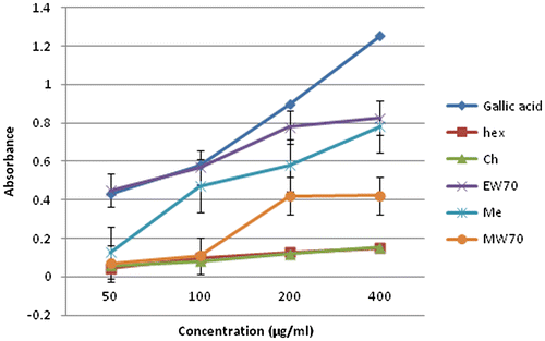
Figure 4. Comparative hydrogen peroxide scavenging activity of Gallic acid Myrothamnus flabellifolius extract.
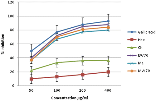
Figure 5. Comparative nitric oxide scavenging activity of Gallic acid Myrothamnus flabellifolius extract.
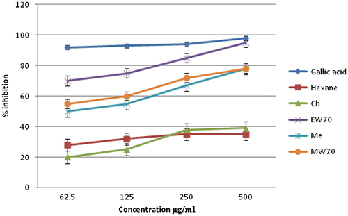
Table 3. Phytochemical screening of Myrothamnus flabellifolius extracts
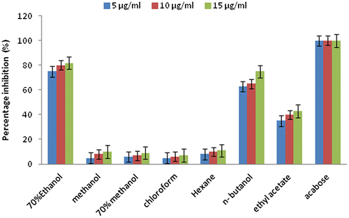
Figure 6. Percentage inhibition of Myrothamnus flabellifolius extracts on α-amylase. Each value is mean ± SEM of three trials.
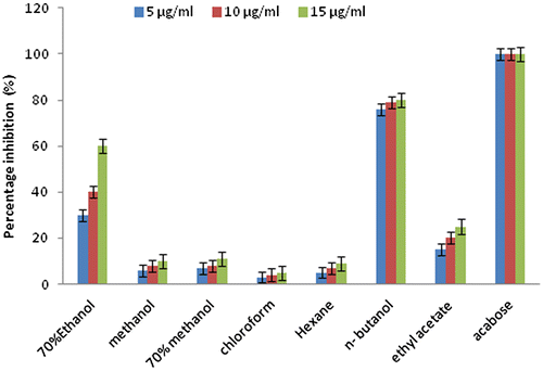
Figure 7. Percentage inhibition of Myrothamnus flabellifolius extracts on α-Glucosidase. Each value is mean ± SEM of three trials.