910
Views
2
CrossRef citations to date
0
Altmetric
Research Article

New evidence for the diversity of mechanisms and protonated Schiff bases formed in the non-enzymatic covalent protein modification (NECPM) of HbA by the hydrate and aldehydic forms of acetaldehyde and glyceraldehyde

, , , & | (Reviewing editor)
Article: 1584955 | Received 25 May 2018, Accepted 05 Feb 2019, Published online: 26 Mar 2019

Figures & data

Figure 1. Structures of salient species in the assessment of the nonenzymatic covalent protein modification of HbA by acetaldehyde or glyceraldehyde, with potential effector reagents 2,3-bisphosphoglycerate (2,3-BPG) and inorganic phosphate (Pi).
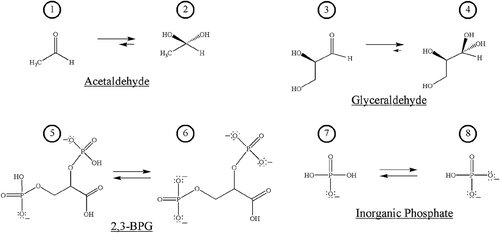
Structures 1 and 2 represent the aldehydic and hydrate forms of acetaldehyde. The relative length of the equilibrium arrows indicate the relative equilibrium concentrations of each form in aqueous solution (33% aldehyde, 66% hydrate). Structures 3 and 4 represent the aldehydic and hydrate forms of glyceraldehyde that exist in equilibrium (4% aldehyde, 96% hydrate). Structures 5 and 6 represent di-anionic and tetra-anionic 2,3-bisphosphoglycerate (2,3-BPG) that exist in equilibrium based upon pH. Structures 7 and 8 represent two forms of inorganic phosphate: mono-anionic Pi, 7, and di-anionic Pi, 8, both of which exist under physiological conditions.
Figure 1. Structures of salient species in the assessment of the nonenzymatic covalent protein modification of HbA by acetaldehyde or glyceraldehyde, with potential effector reagents 2,3-bisphosphoglycerate (2,3-BPG) and inorganic phosphate (Pi).

Figure 2. Generic scheme for the nonenzymatic covalent protein modification (NECPM) of a protein by a non-glucose binding species.

The semi-circle represents a generic protein pocket in which a non-glucose species binds. Here the scheme represents a protein pocket that includes the N-terminus. The NECPM process can proceed in internal protein pockets as well. Once bound in the pocket, the binding species, serving as an electrophile, can undergo nucleophilic attack by a lone pair on an amine group of an amino acid residue (shown here as an N-terminus amino acid) which ultimately results in covalent attachment to the protein via variable mechanisms.
Figure 2. Generic scheme for the nonenzymatic covalent protein modification (NECPM) of a protein by a non-glucose binding species.

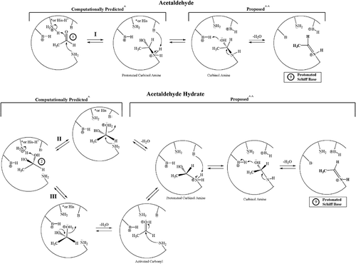
Figure 3. Potential NECPM mechanisms for covalent modification of Val1 of HbA that are geometrically possible for acetaldehyde 1 (Mechanism I) and the acetaldehyde hydrate 2 (Mechanisms II and III) based upon molecular modeling with MOE.1
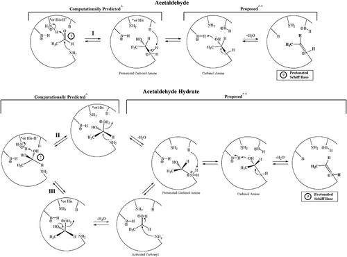
These mechanisms were predicted to be geometrically able to proceed based upon energetic minima calculated from MOE. In the top box, mechanism I, species 1 binds and then the nucleophilic lone pair on the nitrogen of a terminal valine amino acid residue attacks the carbonyl carbon of 1. This causes the π-bond of the carbonyl to break, enhancing the basicity of the carbonyl oxygen, which then abstracts a proton from a nearby acidic amino acid residue (Lys NH3+ or a protonated histidine). This forms the protonated carbinol amine. A basic amino acid residue can then deprotonate the amine, generating the carbinol amine. The lone pair on the nitrogen can then form a π-bond, as the alcohol group (hydoxyl group) abstracts a proton from a nearby acidic amino acid residue and departs as water. This forms the protonated Schiff base, 9. Mechanisms II and III begin with the hydrate form of acetaldehyde, 2, bound in the protein pocket. In Mechanism II, the oxygen of an alcohol group on 2 abstracts a proton from a nearby acidic amino acid residue. The lone pair on the nitrogen of a terminal valine then attacks as a nucleophile, causing water to depart as a leaving group. This generates the protonated carbinol amine. In Mechanism III, the oxygen of an alcohol group on 2 abstracts a proton from a nearby acidic amino acid residue. The lone pair on the oxygen of the remaining alcohol group of 2 forms a π-bond to carbon. This facilitates the loss of water as a leaving group and generates an activated carbonyl species. The carbonyl carbon is then attacked by a nucleophilic lone pair on the nitrogen of a terminal valine to form the protonated carbinol amine. Both Mechanisms II and III involve an identical protonated carbinol amine that leads to the formation of the same protonated Schiff base, 9.
Figure 3. Potential NECPM mechanisms for covalent modification of Val1 of HbA that are geometrically possible for acetaldehyde 1 (Mechanism I) and the acetaldehyde hydrate 2 (Mechanisms II and III) based upon molecular modeling with MOE.1

Table 1. Computational modeling of acetaldehyde (aldehyde 1 and hydrate 2) binding in the HbA1c pocket of human hemoglobin as both single-species binding and concomitant binding with potential effector reagents, 2,3-BPG (5 or 6) or Pi (7 or 8)

Table 2. Computational modeling of Glyceraldehyde/Hydrate binding to the HbA1c pocket of human hemoglobin both as single-species binding and with concomitant binding with 2,3-BPG or with Pi

Figure 4. Potential NECPM mechanisms for HbA covalent modification that are geometrically possible for glyceraldehyde 3 (Mechanism I) and the glyceraldehyde hydrate 4 (Mechanisms II and III) based upon molecular modeling with MOE.2
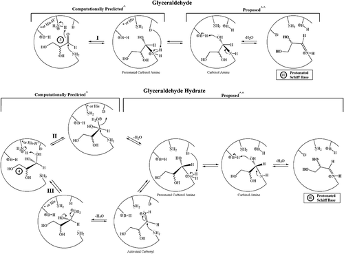
These three mechanisms are predicted to be geometrically feasible based upon calculated energetic minima from computations conducted in MOE. In Mechanism I, the aldehydic form of glyceraldehyde, 3, binds and the nucleophilic lone pair on the nitrogen of a terminal valine amino acid residue attacks the carbonyl carbon of 3. This causes the π-bond to break and enhances the basicity of the carbonyl oxygen, which then abstracts a proton from a nearby acidic amino acid residue (Lys NH3+ or a protonated histidine). This generates the protonated carbinol amine. Once formed, a basic amino acid residue can deprotonate the positively charged amine to form the carbinol amine. The lone pair on nitrogen can then form a π-bond, increasing the basicity of the oxygen of the alcohol which can then abstract a proton from an acidic amino acid reside. The protonated alcohol group then departs as water. This forms the protonated Schiff base, 10. Mechanisms II and III begin with the hydrate form of glyceraldehyde, 4, bound in the protein pocket. In Mechanism II, the oxygen of an alcohol group on 4 abstracts a proton from a nearby acidic amino acid residue. The lone pair on the nitrogen of a terminal valine then attacks as a nucleophile, causing water to depart as a leaving group. This generates the protonated carbinol amine. In Mechanism III, the oxygen of an alcohol group on 4 abstracts a proton from a nearby acidic amino acid residue. The lone pair on the oxygen of the remaining alcohol group of 4 forms a π-bond to carbon. This facilitates the loss of water as a leaving group and generates an activated carbonyl species. The carbonyl carbon is then attacked by a nucleophilic lone pair on the nitrogen of a terminal valine to form the protonated carbinol amine. Both Mechanisms II and III involve a common protonated carbinol amine species and the following chemistry from the common intermediate leads to the formation of the same protonated Schiff base, 10.
Figure 4. Potential NECPM mechanisms for HbA covalent modification that are geometrically possible for glyceraldehyde 3 (Mechanism I) and the glyceraldehyde hydrate 4 (Mechanisms II and III) based upon molecular modeling with MOE.2
Supplemental material

Supplemental Material

Download MS Word (21.7 KB)