Figures & data
Figure 1. (a–i) The structure of the plant, inflorescence, and fruit of M. pomifera at different developmental stages. (a) Whole plant morphology; (b,c) Developing inflorescences with short peduncles on a branch of the plant, bracts at the base of the inflorescences and the styles on the inflorescence surface can be seen; (d–g) Continuation of inflorescence and styles development and multiple accessory fruit formation; styles change from felt-like to needle-like and some of them are dried and shed. (h) Transverse section of multiple accessory fruit with multiple distinguishable flowers, a few number of filled seeds are formed. (i) Drupe fruits with long and permanent styles.
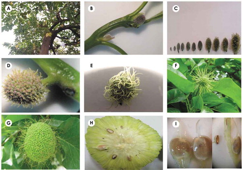
Figure 2. Anatomical structure of inflorescence in longitudinal (a) and transverse (b) views. The structure of peduncle, grown receptacle and flowers without pedicle are seen (PF: pistillate flower; RE: receptacle; PE: peduncle).
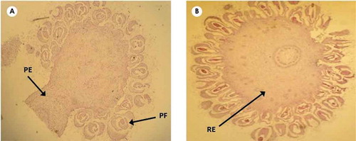
Figure 3. (a–h) The structure and development of flower, pistil and ovule. (a) Transverse section of inflorescence with flowers containing tetrameric and developing perianths. (b) Pistil structure in longitudinal view, perianth, developing ovule and style in its upper part are obvious. (c–h) Apical initiation of ovule, its rotation and formation of tenuinucellate, ana-campylotropous ovule, megaspore mother cell is differentiated in ovule, the development of perianth and formation of secretory and alveolar structures in (g) and (h) are seen (MM: megaspore mother; OV: ovule; Pi: pistil; OP: ovule primordium; Pe: perianth; SS: secretory structure; Mi: micropyle; St: style; IN: inner integument; OU: outer integument).
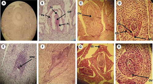
Figure 4. (a–e) The development of embryo sac and embryo. (a,b) Ana–campylotropous ovule with apical placentation and 90 degrees rotation upwards and pear-shaped embryo sac. (c–e) Developmental stages of embryo including globular, heart-shaped. Short multi-cellular suspensor is seen in (d) and (e).
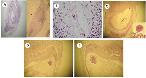
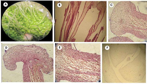
Figure 5. (a–g) Fruit structure in the cotyledonary and curved embryo stage. (a) Cotyledonary embryo in the early stages, the volume of endosperm is still high. (b–d) Curved embryo; fruit pericarp and style are obvious in (b) and (c). Shoot apical meristem (SAM) in (d) and root apical meristem (RAM) in (e) and (f) are observed. In (f), the separation of fruit pericarp from seed integument (brown structure on endosperm with 3–5 cell layers) is seen.
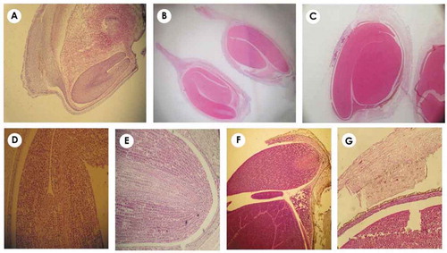
Figure 6. (a–d) The morphology of true fruit, seed and embryo in transverse, longitudinal and diagonal sections. The fruit exo-mesocarp and endocarp are fleshy and stony, respectively, showing drupe (drupelet) appearance. Style is permanent and seed coat surrounding endosperm is seen brown color.
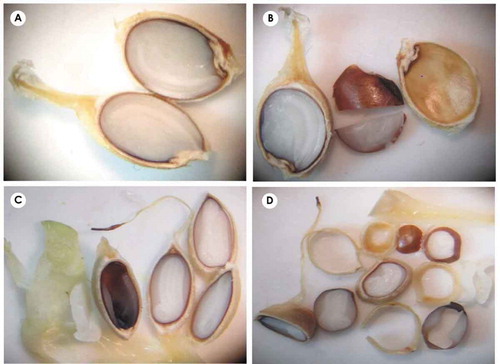
Figure 7. (a–f) Morphological and anatomical structure of pome like and false fruit and its simple drupelets. (a) Pome like fruits with styles located in the center of each drupelet. (b–e) Longitudinal view of perianth with grown and globular apex; the elongated and detached cells located in the middle of perianth are secretory. (f) Perianth structure and its secretory cells in the transverse view, simple fruit is observed in the centre of perianth.