
Figures & data
Figure 1. Preoperative picture showing the defect in the upper lip and the large laceration of the lower lip.
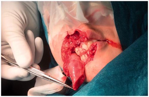
Figure 2. The amputated part of the upper lip, measuring 4.5 × 3 cm, was kept in a hypothermic state.
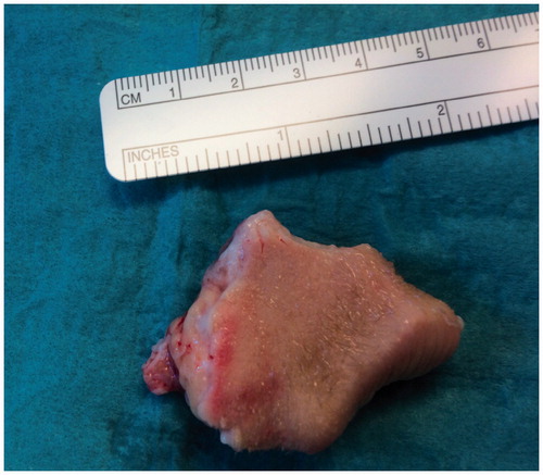
Figure 3. Shortly after the lip was reapproximated, venous congestion occurred. A leech was immediately applied and the lip regained normal color.
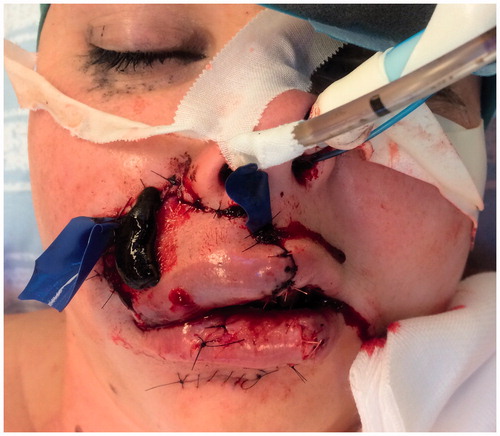
Figure 4. The patient at one year follow-up. The lip still has a slight edema, which is slowly subsiding.
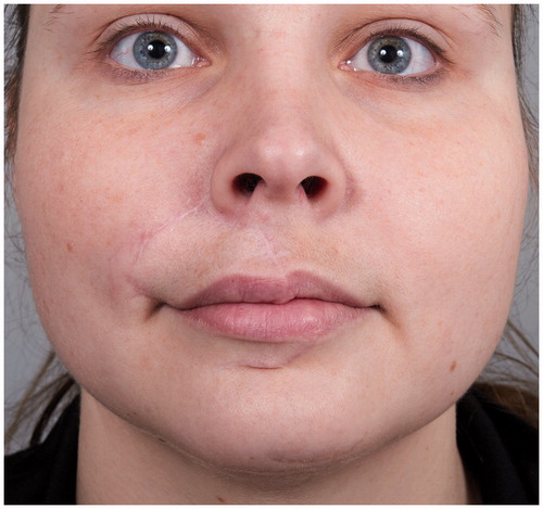
Table 1. Lip replantation cases reported in English literature (n = 32).