Figures & data
Figure 1. (a) Eight years before surgical excision (17 × 12 × 14 mm). (b) Four years before surgical excision (28 × 18 × 30 mm). (c) Preoperative (60 × 20 × 35 mm).
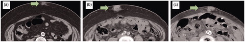
Figure 2. (a) Preoperative appearance. (b) The excised mass with indurated subcutaneous fat.
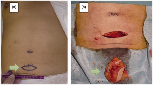
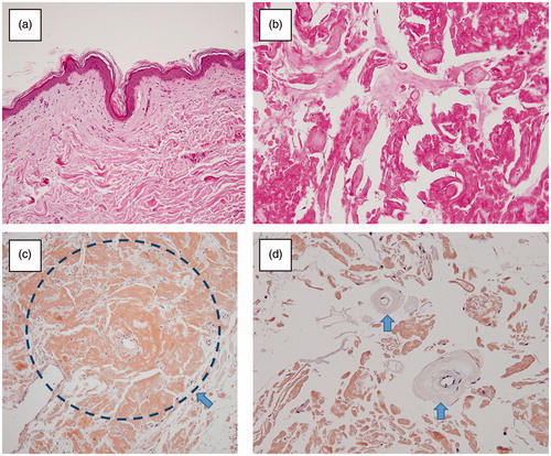
Figure 3. (a,b) The subcutaneous tissue was degenerated broadly (Haematoxylin and eosin staining). (c) Extensive amyloid deposition in the subcutaneous tissue (arrow) (Congo red staining). (d) No evidence of vascular involvement (arrow: subcutaneous artery without amyloid deposition) (Congo red staining).