Figures & data
Figure 1. CT images showing an irregular articular surface due to poor reduction in the initial surgery.
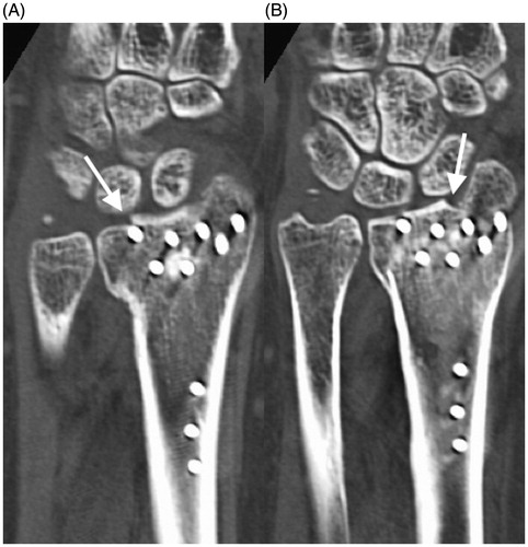
Figure 2. (A) Immediately postoperative radiograph of the radiolunate arthrodesis with the iliac crest. (B) Radiograph at 6 months after the first radiolunate arthrodesis showing nonunion.
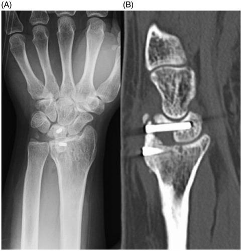
Figure 3. (A) Intraoperative photograph of the vascularized bone graft at the time of harvesting. (B) Immediately postoperative photograph of the wrist with the external fixation device. (C) At 3 months after the last surgery, a radiograph confirmed bony union.
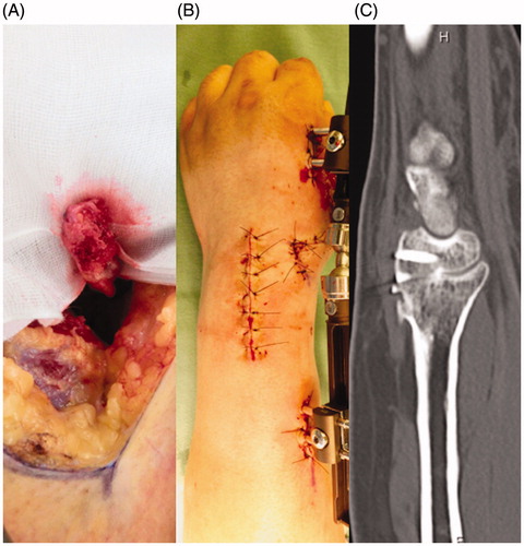
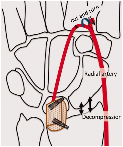
Figure 4. Diagrams of the surgical procedure. The bone graft was placed between the lunate and radius with a slight distraction of the lunate. End–to-end anastomosis was performed into the dorsal branch of the radial artery.