Figures & data
Figure 1. On initial presentation, the patient had a 1 cm slightly bluish, pulsatile, non-tender soft tissue mass over the course of the left radial artery, concerning for a vascular anomaly.
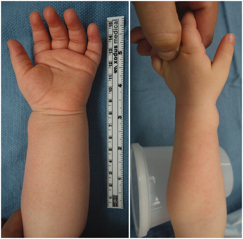
Figure 2. Magnetic resonance angiography (coronal, sagittal, and axial views) revealed a 1.1 × 0.8 × 1.0 cm mass with retrograde arterial filling arising laterally off the distal radial artery from the superficial palmar arch with evidence of thrombosis of the artery proximal to the lesion and intact superficial and deep palmar arches.
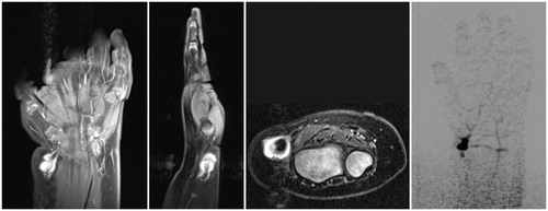
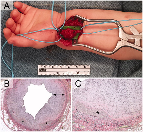
Figure 3. A. Intraoperatively, the lesion was pulsatile, grossly multi-lobular, and reddish and focally bluish in colour with fibrous attachments to the flexor carpi radialis sheath. The radial artery was resected proximally to the mid-forearm and distally just past the wrist flexion crease. Photomicrographs exhibiting pseudoaneurysmal radial artery with scant residual media (asterisks) and striking intimal fibroplasia (arrow). Verhoeff-van Giesson stains (3B) original magnification, ×40. (3C) original magnification, ×500.