
Figures & data
Figure 1. Entrance wound on the radial aspect of the right long finger middle phalanx.
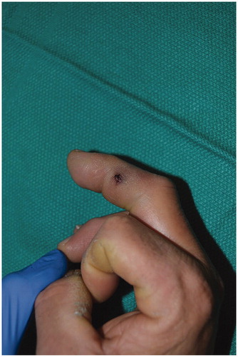
Figure 2. Radiograph demonstrating foreign material in the volar aspect of the right long finger, extending far beyond the injection site, lateral (left) and AP (right) views.
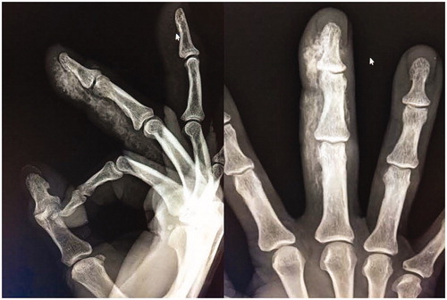
Figure 3. (left) Operative exploration with exposure of the neurovascular bundles and flexor sheath of the finger. The fresh white paint is clearly visible; (right) After extensive washout and initial debridement.
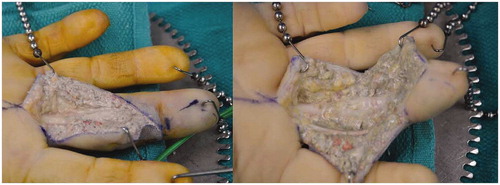
Table 1. Number of cases of each type of injected material.
Table 2. Rate of complications of each case and the resulting outcomes.