Figures & data
Figure 1. Inches x-ray: calcification of LCL of MCPP.
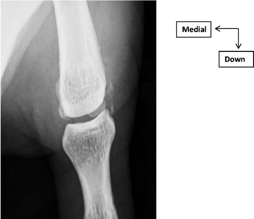
Figure 2. Thumb ultrasound: calcified appearance of MCPL LCL with synovial hypertrophy.
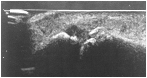
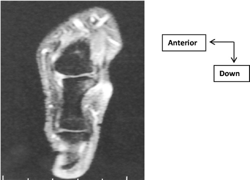
Figure 3. MRI: (sagittal section fat sat T2) Thickened aspect of LCL of MCPP which is in Hypo-signal T1 Hypo-signal T2.