Figures & data
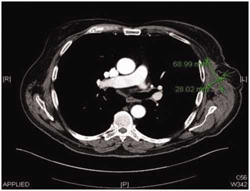
Figure 1. Preoperative CT scan image showing left axillary chronic seroma.
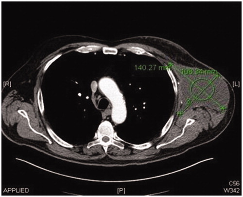
Figure 2. Preoperative left lateral view of the patient showing the presence of the large left axillary encapsulated seroma.
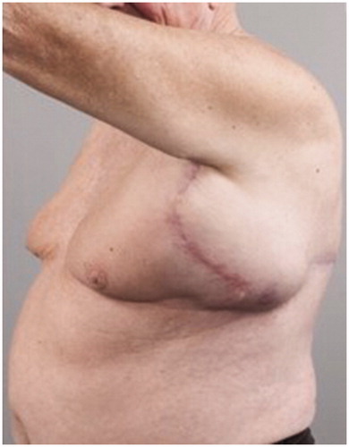
Figure 3. Preoperative posterior view of the patient showing the presence of the large left axillary encapsulated seroma and the malignant melanoma scar.
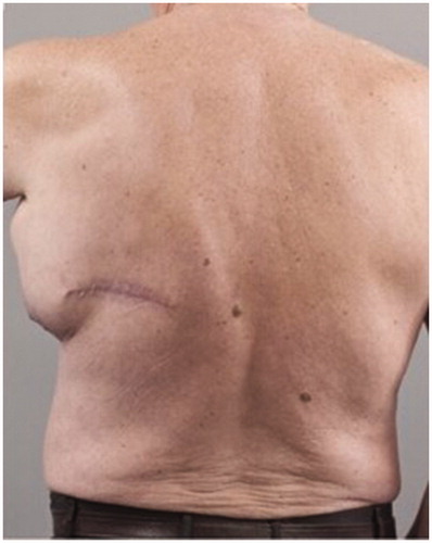
Figure 4. Post resection Intraoperative view of the large encapsulated seroma.
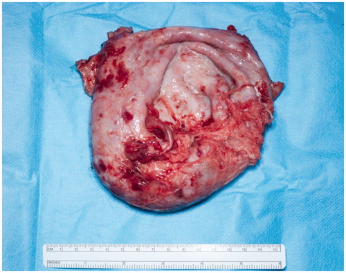
Figure 5. ALT flap inset in the axillary area to obliterate dead space.
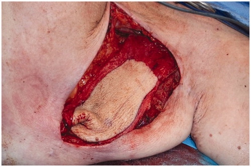
Figure 6. CT scan image 1-month post-resection of chronic seroma capsule and free ALT-flap inset.
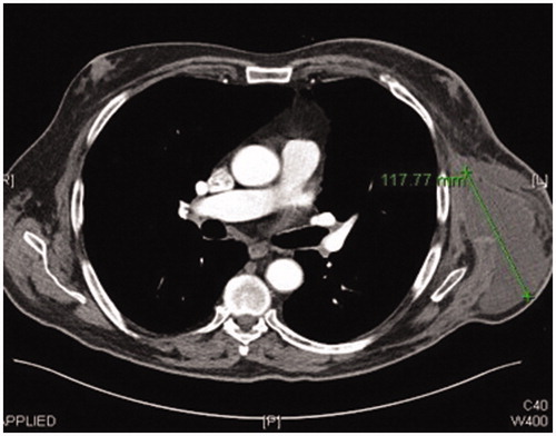
Figure 7. CT scan image 3-month post-resection of chronic seroma capsule showing almost complete resolution.