Figures & data
Figure 1. (A, B) Preoperative appearance: marked ptosis and crepey eyelid skin.
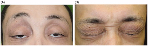
Figure 2. (A) Palpebral skin exhibits epidermal spongiosis and perivascular lymphocyte infiltration, suggesting superficial dermatitis compatible with blepharochalasis (hematoxylin and eosin, ×200). (B) Elastic fibers are reduced in the superficial dermis and exhibit fragmentation (arrowhead, elastica van Gieson, ×200). (C) Immunostaining reveals perivascular IgA deposits (arrow, ×100).
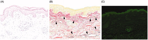
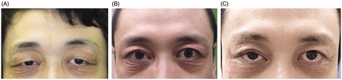
Figure 3. (A) Appearance at 3 months after the first operation. (B) Appearance at 8 months after the second operation. (C) Appearance at 15 months after the third operation.