
Figures & data
Figure 1. Clinical findings and MRI images. (A) A reddish mass of 4-mm diameter is seen under the nail plate (area surrounded by the triangle mark). Distal nail splitting is observed in the left thumb. (B) T1-weighed magnetic resonance image shows tumor with normal intensity. (C) T2-weighed magnetic resonance image shows high-intensity lesion. Flow void is indicated by an arrow. MRI: magnetic resonance imaging.
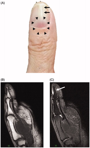
Figure 2. Surgical findings. (A) A reddish mass is seen under the nail plate. The excision line after the nail claw is indicated in blue. (B) After excision, skin grafting was performed from the thenar eminence.
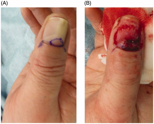
Figure 3. Histopathological findings. (A) A loupe image. An incompletely encapsulated tumor. (B) A middle-power view. The tumor is composed of fine collagen fibers pointed in every direction. (C) A high-power view. Nuclei of the proliferating cells are spindle- or comma-shaped. Among the tumor cells, capillaries and a small number of mast cells are dispersed (indicated by arrows). (D) Appearance of new thumb nail six months postoperatively. This observation is natural.
