Figures & data
Figure 1. A 62-year-old Indian male with a cT4aN1M0 intraoral lesion. (A) Exophytic ulcerative squamous cell carcinoma of the left buccal mucosa. (B) Left side of panoramic dental x-ray demonstrating absence of bony invasion. This finding was corroborated by CT scan. (C–D) CT neck revealed a 2.3 × 1.9 × 2.3 cm tumor at the left buccal mucosa and a 2.3 × 1.3 cm centrally necrotic ipsilateral lymph node at level 1B.
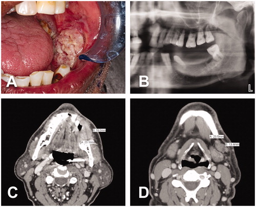
Figure 2. Intraoperative views. (A-B) Marking of the oncological skin excision on the left cheek and design of the large medially-based left cervicothoracic rotation flap. (C–D) Cervicothoracic flap already harvested and reflected medially. Completion of tumor resection (including surrounding tissues and cheek skin), segmental mandibulectomy and left modified radical neck dissection.
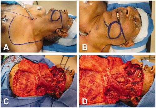
Figure 3. Intraoperative views (cont’d). (A) Surgical specimen including tumor and surrounding tissues, cheek skin, floor of mouth, segment of left mandible and lymphoareolar tissues of the left modified radical neck dissection. (B) Advancement and rotation of the cervicothoracic flap to cover the cheek defect. (C–D) Inset of the cervicothoracic flap.
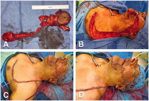
Figure 4. Donor site of osteocutaneous radial forearm free flap. (A) Use of reconstruction plate to protect radius bony defect after flap harvest. Two distal bicortical and three proximal bicortical screws were placed. (B) Intraoperative view of donor site of left forearm. (C) Long-term follow-up.
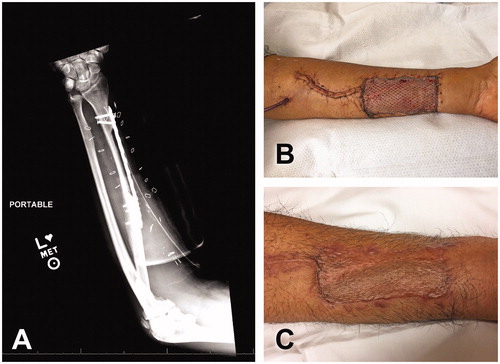
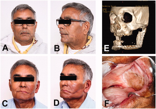
Figure 5. Long-term follow-up. (A-B) Preoperative pictures for evaluation of the surgical outcome. (C-D) Frontal and oblique views fourteen months after surgery. Patient has completed radiation therapy. (E) Image of CT facial bones 3D reconstruction with satisfactory osseous alignment between the bony segment of radius and remaining mandible with no blast or lytic process. (F) Intraoral image showing the left buccal vestibule and its lining with the skin paddle of radial forearm free flap.