Figures & data
Figure 1. Top: Right posterior parietal scalp with skin lesion. Bottom left: Closer-up view of lesion and scalp. Bottom right: Post-excision as well as showing inferior biopsy site.
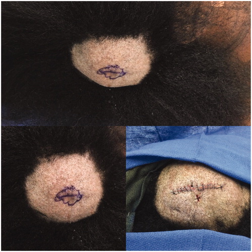
Figure 2. The tumor consists of lobules of basaloid to multivacuolated tumor cells, the latter containing clear vacuoles in the cytoplasm, indicating sebaceous differentiation. Top left: 100x magnification. Bottom left and Top right: 50x magnification. Bottom right: 200x magnification.
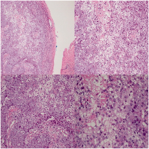
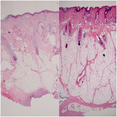
Figure 3. Left: pathology slide showing normal skin of the scalp. Right: pathology slide showing the thicker specimen, increased adipose tissue and edematous aspect of the lipedematous scalp.