Figures & data
Figure 1. (A and B) Pre-operative x-rays of the first case.
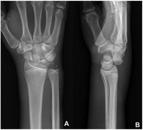
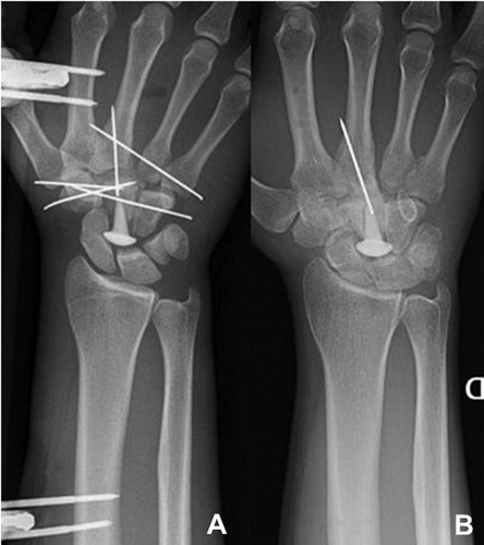
Figure 2. (A) X-ray after the first surgery. Note the empty space at the place of the proximal pole of the capitate. (B) X-ray after the second surgery, the K-wires were replaced by the RCPI prosthesis and a screw was inserted in the scaphoid. (C) At 21 months’ follow-up (all case 1).
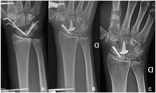
Figure 3. Pre-operative CT-scan case 2.
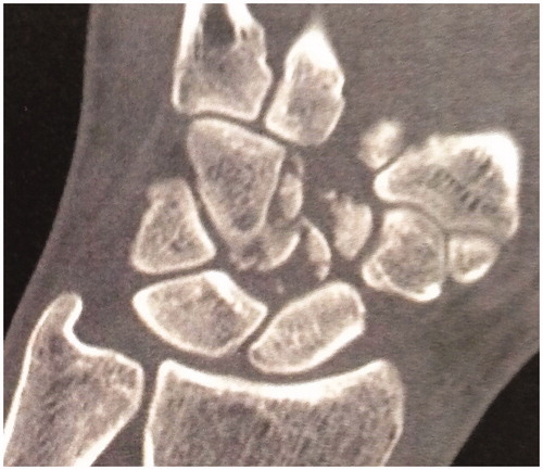
Figure 4. (A) Post-operative x-ray case 2. (B) X-ray at 29 months’ follow-up, after removal of the external fixation and K-wires.