Figures & data
Table 1. Cases of B-cell lymphomas associated with breast implants.
Figure 1. The patient’s abdominal CT scan demonstrates massive splenomegaly with adjacent abdominal lymphadenopathy.
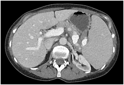
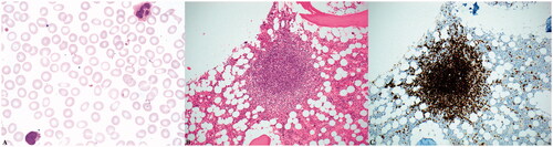
Figure 2. Microscopic examination of the patient’s peripheral blood smeared reveals atypical lymphocytes with villous morphology (A, 1000x magnification). Her bone marrow contained prominent lymphoid aggregates accounting for approximately 20% of the marrow’s cellularity (B, 200x magnification) that were composed of B-cells positive for CD20 by immunohistochemistry (C, 200x magnification).