
Figures & data
Figure 1. A small wound is observed on the thenar aspect of the thumb, and the paint is attached to the surrounding wound.
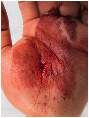
Figure 2. X-ray (a, b) and computed tomography (c, d) images show that the paint spread extensively along the flexor tendons of the palm.
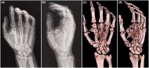
Figure 3. (a) Paint is observed inside the carpal tunnel (arrow: paint, dashed arrow: median nerve). (b) There is also paint on the dorsal side of the proximal phalanx (arrow). (c) After removing paint, the median nerve is evaluated (arrow: median nerve).
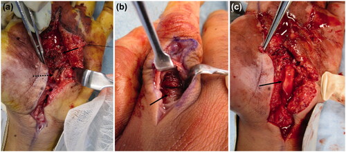
Figure 4. Computed tomography images acquired after the first operation show paint in the thenar space and the index, middle, and ring fingers.
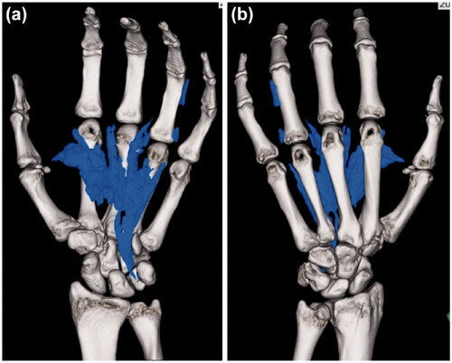
Figure 5. (a) Clay-like adherent paint is observed in the thenar space (arrow). (b) After removing paint, the inside of the carpal tunnel was checked, and paint is observed between the flexor tendons (solid arrow: palmer aponeurosis, dashed arrow: paint).
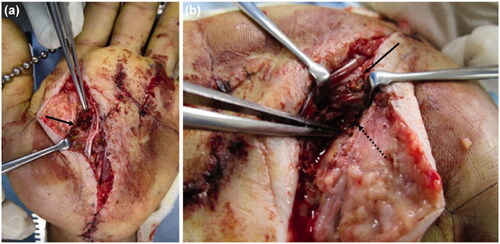
Figure 6. (a) Extensive necrosis is observed around the wound. (b) Debridement of the necrotic epidermis reveals residual paint in the carpal tunnel. (c) The extracted paint is shown. A large amount of cured paint was also observed during the third operation. (d) After debridement, a fan-shaped skin defect is seen in the hand. (e) Reconstruction was performed with a pedicled groin flap.
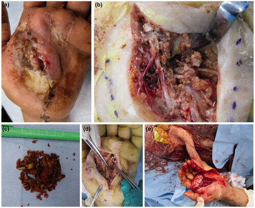
Figure 7. (a–d) No limitation in the ROM of the fingers is observed at the latest follow-up.
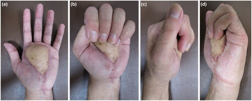
Table 1. Total active motion of the metacarpophalangeal joints.