Figures & data
Figure 1. Axial MRI T1 weighted image with Fast Spin Echo (FSE) showing hyperintense lesion on both the volar and dorsal side of ring finger.
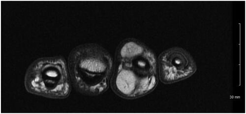
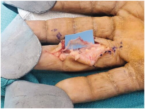
Figure 2. Intra operative image showing the multilobulated tumor.
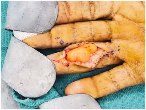
Figure 3. Dorsal extension of the tumor.
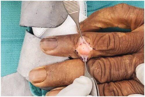
Figure 4. Excised specimen. The mass was divided at the middle (Arrow) .
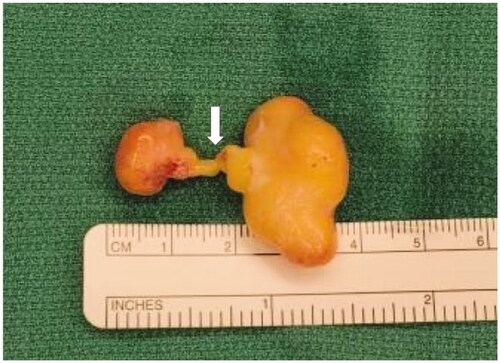
Figure 5. The intact nerve after excision of the tumor.